 Explore
 Validate
 Learn
Learn Western blot
Antibody data
- Antibody Data
- Antigen structure
- References [0]
- Comments [0]
- Validations
- Western blot [1]
- Immunocytochemistry [1]
- Immunohistochemistry [1]
Submit
Validation data
Reference
Comment
Report error
- Product number
- TA328654 - Provider product page

- Provider
- OriGene
- Product name
- Rabbit Polyclonal Anti-TRPV3 (extracellular)
- Antibody type
- Polyclonal
- Description
- Rabbit Polyclonal Anti-TRPV3 (extracellular)
- Host
- Rabbit
- Conjugate
- Unconjugated
- Epitope
- TRPV3
- Antibody clone number
- NULL
- Vial size
- 200 µl
- Concentration
- NULL
No comments: Submit comment
Supportive validation
- Submitted by
- OriGene (provider)
- Main image
- Experimental details
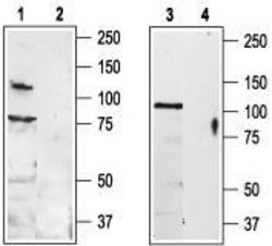
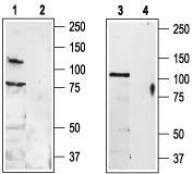
- Western blot analysis of rat DRG (lanes 1 and 2) and rat brain (lanes 3 and 4) lysates: 1, 3. Anti-TRPV3 (extracellular) antibody, (1:200). 2, 4. Anti-TRPV3 (extracellular) antibody, preincubated with the control peptide antigen.
- Validation comment
- WB
Supportive validation
- Submitted by
- OriGene (provider)
- Main image
- Experimental details
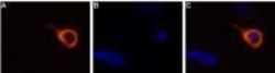
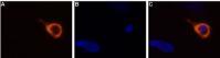
- Expression of TRPV3 in rat DRG primary culture. Immunocytochemical staining of living rat dorsal root ganglion (DRG) primary culture using Anti-TRPV3 (extracellular) antibody, (1:50-1:100), followed by goat anti-rabbit-AlexaFluor-555 secondary antibody (A). B. Nuclear staining with the cell-permeable dye Hoechst 33342. C. Merged image of panels A and B.
- Validation comment
- IF
Supportive validation
- Submitted by
- OriGene (provider)
- Main image
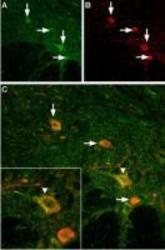
- Experimental details
- IHC staining of mouse spinal cord using Anti-TRPV3 (extracellular) antibody. A. TRPV3 (green) appears in neurons (vertical arrows) in the ventral horn of the mouse spinal cord. B. Motor neurons were stained with goat anti choline-acetyltransferase (red). In some motor neurons (A) TRPV3 was more intense (vertical arrow) whereas in others it was weak (horizontal arrows). C. Merged images of panels A and B. The inset in C magnifies one large motor neuron (vertical arrowhead).
- Validation comment
- IHC
