Explore
 Validate
 Learn
Learn Western blot
Antibody data
- Antibody Data
- Antigen structure
- References [0]
- Comments [0]
- Validations
- Western blot [1]
- Immunocytochemistry [1]
- Immunohistochemistry [1]
Submit
Validation data
Reference
Comment
Report error
- Product number
- OAAB16995 - Provider product page

- Provider
- Aviva Systems Biology
- Product name
- DCAMKL1 antibody - C - terminal region
- Antibody type
- Polyclonal
- Reactivity
- Human, Mouse
- Host
- Rabbit
- Vial size
- 400ul
- Storage
- Maintain refrigerated at 2-8°C for up to 6 months. For long term storage store at -20°C in small aliquots to prevent freeze-thaw cycles.
No comments: Submit comment
Supportive validation
- Submitted by
- Aviva Systems Biology (provider)
- Main image
- Experimental details
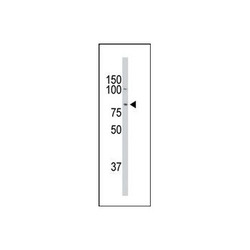
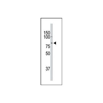
- The DCAMKL1 Antibody (C-term ) (Cat. # OAAB16995) western blot analysis in mouse heart tissuelysates (35µg/lane). This demonstrates the DCAMKL1 antibody detected the DCAMKL1 protein (arrow).
Supportive validation
- Submitted by
- Aviva Systems Biology (provider)
- Main image
- Experimental details
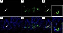
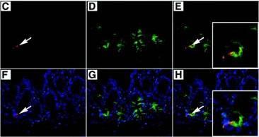
- (C): The cell positive for DCAMKL-1, stained red (indicated by the arrow) , appears at the base of the crypt. (D): Intestinal section stained for Musashi-1 (green). (E): Colocalization of DCAMKL-1 and Musashi-1 (yellow, indicated by the arrow). The magnified inset image represents the single cell positive for both DCAMKL-1 and Musashi-1. (F): Costaining of DCMAKL-1 (red, indicated by the arrow) with nuclear Hoechst 33342 (blue) staining. (G): Costaining of Musashi-1 (green) with nuclear Hoechst 33342 (blue) staining. (H): Colocalization of DCAMKL-1 and Musashi-1 (yellow, indicated by the arrow) , costained with nuclear Hoechst 33342 (blue) staining. The magnified inset image represents the single cell positive for both DCAMKL-1 and Musashi-1 (yellow) .
Supportive validation
- Submitted by
- Aviva Systems Biology (provider)
- Main image

- Experimental details
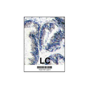
- Formalin-fixed and paraffin-embedded human hepatocarcinoma reacted with DCAMKL1 Antibody (C-term) (Cat.# OAAB16995) , which was peroxidase-conjugated to the secondary antibody, followed by DAB staining. This data demonstrates the use of this antibody for immunohistochemistry; clinical relevance has not been evaluated.