 Explore
 Validate
 Learn
Learn Western blot
 ELISA
ELISAAntibody data
- Antibody Data
- Antigen structure
- References [0]
- Comments [0]
- Validations
- Western blot [1]
- Immunohistochemistry [2]
- Flow cytometry [1]
Submit
Validation data
Reference
Comment
Report error
- Product number
- STJ98362 - Provider product page

- Provider
- St John's Laboratory
- Product name
- Anti-RICTOR antibody [7B3] (STJ98362)
- Antibody type
- Monoclonal
- Description
- Mouse monoclonal antibody anti-Rapamycin-Insensitive Companion Of Mtor is suitable for use in Western Blot, Immunohistochemistry, Immunofluorescence, Immunocytochemistry, Flow Cytometry and ELISA research applications.
- Reactivity
- Human, Mouse, Simian
- Host
- Mouse
- Conjugate
- Unconjugated
- Antigen sequence
NA- Epitope
- NA
- Isotype
- IgG
- Antibody clone number
- NA
- Vial size
- NA
- Concentration
- NA
- Storage
- Store at-20°C for up to 1 year from the date of receipt, and avoid repeat freeze-thaw cycles.
- Handling
- NA
No comments: Submit comment
Supportive validation
- Submitted by
- St John's Laboratory (provider)
- Main image

- Experimental details
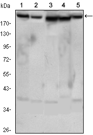
- Western blot analysis using Rictor monoclonal antibody against HeLa (1) , PANC-1 (2) , MOLT4 (3) , HepG2 (4) and HEK293 (5) cell lysate.
- Sample type
- NA
- Validation comment
- NA
- Primary Ab dilution
- NA
- Other comments
- NA
- Secondary Ab
- NA
- Secondary Ab dilution
- NA
- Protocol
- NA
Supportive validation
Supportive validation
- Submitted by
- St John's Laboratory (provider)
- Main image
- Experimental details
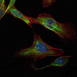
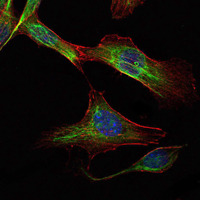
- Immunofluorescence analysis of NIH/3T3 cells using Rictor monoclonal antibody (green). Blue: DRAQ5 fluorescent DNA dye. Red: Actin filaments have been labeled with Alexa Fluor-555 phalloidin.
- Sample type
- NA
- Validation comment
- NA
- Primary Ab dilution
- NA
- Other comments
- NA
- Secondary Ab
- NA
- Secondary Ab dilution
- NA
- Protocol
- NA
Supportive validation
- Submitted by
- St John's Laboratory (provider)
- Main image
- Experimental details
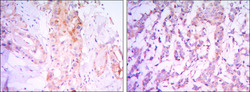
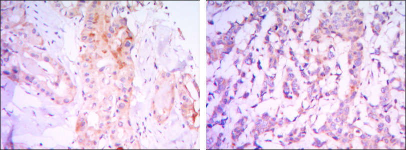
- Immunohistochemistry analysis of paraffin-embedded thyroid gland tissues (left) and human breast carcinoma (right) with DAB staining using Rictor monoclonal antibody.
- Sample type
- NA
- Validation comment
- NA
- Primary Ab dilution
- NA
- Other comments
- NA
- Secondary Ab
- NA
- Secondary Ab dilution
- NA
- Protocol
- NA
Supportive validation
- Submitted by
- St John's Laboratory (provider)
- Main image

- Experimental details
- Flow cytometric analysis of Hela cells using Rictor monoclonal antibody (green) and negative control (purple).
- Sample type
- NA
- Validation comment
- NA
- Primary Ab dilution
- NA
- Other comments
- NA
- Secondary Ab
- NA
- Secondary Ab dilution
- NA
- Protocol
- NA
