 Explore
 Validate
 Learn
Learn Western blot
 ELISA
ELISA Immunocytochemistry
Antibody data
- Antibody Data
- Antigen structure
- References [2]
- Comments [0]
- Validations
- Immunocytochemistry [5]
- Immunohistochemistry [1]
- Flow cytometry [2]
Submit
Validation data
Reference
Comment
Report error
- Product number
- MA5-15681 - Provider product page

- Provider
- Invitrogen Antibodies
- Product name
- RICTOR Monoclonal Antibody (7B3)
- Antibody type
- Monoclonal
- Antigen
- Purifed from natural sources
- Description
- MA5-15681 targets RICTOR in indirect ELISA, FACS, IF, IHC, and WB applications and shows reactivity with Human, mouse, and Non-human primate samples. The MA5-15681 immunogen is purified recombinant fragment of human RICTOR expressed in E. Coli. MA5-15681 detects RICTOR which has a predicted molecular weight of approximately 192kDa.
- Reactivity
- Human, Mouse
- Host
- Mouse
- Isotype
- IgG
- Antibody clone number
- 7B3
- Vial size
- 100 μL
- Concentration
- Conc. Not Determined
- Storage
- Store at 4°C short term. For long term storage, store at -20°C, avoiding freeze/thaw cycles.
Submitted references Lamellipodin-RICTOR Signaling Mediates Glioblastoma Cell Invasion and Radiosensitivity Downstream of EGFR.
Folliculin regulates mTORC1/2 and WNT pathways in early human pluripotency.
Moritz S, Krause M, Schlatter J, Cordes N, Vehlow A
Cancers 2021 Oct 24;13(21)
Cancers 2021 Oct 24;13(21)
Folliculin regulates mTORC1/2 and WNT pathways in early human pluripotency.
Mathieu J, Detraux D, Kuppers D, Wang Y, Cavanaugh C, Sidhu S, Levy S, Robitaille AM, Ferreccio A, Bottorff T, McAlister A, Somasundaram L, Artoni F, Battle S, Hawkins RD, Moon RT, Ware CB, Paddison PJ, Ruohola-Baker H
Nature communications 2019 Feb 7;10(1):632
Nature communications 2019 Feb 7;10(1):632
No comments: Submit comment
Supportive validation
- Submitted by
- Invitrogen Antibodies (provider)
- Main image
- Experimental details
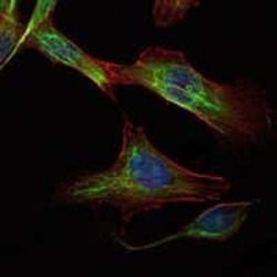
- Immunofluorescence analysis of NIH/3T3 cells using RICTOR monoclonal antibody (Product # MA5-15681) (Green). Blue: DRAQ5 fluorescent DNA dye. Red: actin filaments have been labeled with phalloidin.

- Submitted by
- Invitrogen Antibodies (provider)
- Main image
- Experimental details
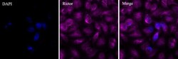
- Immunofluorescent analysis of Rictor (magenta) in Hela cells. The cells were fixed with 4% paraformaldehyde for 15 minutes, permeabilized with 0.1% Triton X-100 in PBS for 15 minutes, and blocked with 2% BSA in PBS for 60 minutes at room temperature. Cells were stained with a Rictor monoclonal antibody (Product # MA5-15681) at a dilution of 1:200 in staining buffer for 3 hour at room temperature, and then incubated with a Goat anti-mouse IgG Secondary Antibody, Alexa Fluor 647 conjugate (Product # A32728) at a dilution of 1:1000 for 1 hour at room temperature (magenta). Nuclei (blue) were counterstained with DAPI dye. Images were taken at 100X magnification. Data courtesy of Thermo Scientific KOL Program.
- Submitted by
- Invitrogen Antibodies (provider)
- Main image

- Experimental details
- Immunofluorescence analysis of NIH/3T3 cells using RICTOR monoclonal antibody (Product # MA5-15681) (Green). Blue: DRAQ5 fluorescent DNA dye. Red: actin filaments have been labeled with phalloidin.

- Submitted by
- Invitrogen Antibodies (provider)
- Main image

- Experimental details
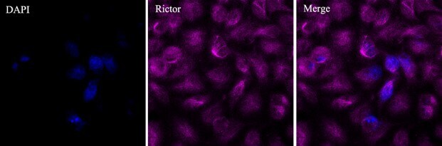
- Immunofluorescent analysis of Rictor (magenta) in Hela cells. The cells were fixed with 4% paraformaldehyde for 15 minutes, permeabilized with 0.1% Triton X-100 in PBS for 15 minutes, and blocked with 2% BSA in PBS for 60 minutes at room temperature. Cells were stained with a Rictor monoclonal antibody (Product # MA5-15681) at a dilution of 1:200 in staining buffer for 3 hour at room temperature, and then incubated with a Goat anti-mouse IgG Secondary Antibody, Alexa Fluor 647 conjugate (Product # A32728) at a dilution of 1:1000 for 1 hour at room temperature (magenta). Nuclei (blue) were counterstained with DAPI dye. Images were taken at 100X magnification. Data courtesy of Thermo Scientific KOL Program.

- Submitted by
- Invitrogen Antibodies (provider)
- Main image
- Experimental details
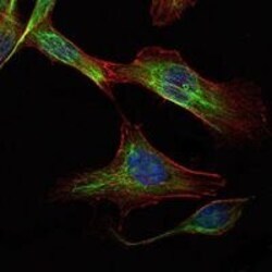
- Immunofluorescence analysis of NIH/3T3 cells using RICTOR monoclonal antibody (Product # MA5-15681) (Green). Blue: DRAQ5 fluorescent DNA dye. Red: actin filaments have been labeled with phalloidin.

Supportive validation
- Submitted by
- Invitrogen Antibodies (provider)
- Main image
- Experimental details
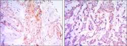
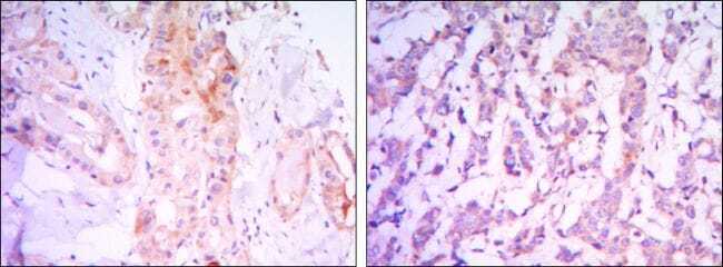
- Immunohistochemical analysis of paraffin-embedded thyroid gland tissues (left) and human breast carcinoma (right) using RICTOR monoclonal antibody (Product # MA5-15681) followed with DAB staining.
Supportive validation
- Submitted by
- Invitrogen Antibodies (provider)
- Main image

- Experimental details
- Flow cytometric analysis of HeLa cells using RICTOR monoclonal antibody (Product # MA5-15681) (green) and negative control (purple).

- Submitted by
- Invitrogen Antibodies (provider)
- Main image

- Experimental details
- Flow cytometric analysis of HeLa cells using RICTOR monoclonal antibody (Product # MA5-15681) (green) and negative control (purple).
