Explore
 Validate
 Learn
Learn Western blot
 Immunocytochemistry
Antibody data
- Antibody Data
- Antigen structure
- References [1]
- Comments [0]
- Validations
- Immunocytochemistry [2]
- Other assay [2]
Submit
Validation data
Reference
Comment
Report error
- Product number
- PA5-101540 - Provider product page

- Provider
- Invitrogen Antibodies
- Product name
- NCAPG Polyclonal Antibody
- Antibody type
- Polyclonal
- Antigen
- Synthetic peptide
- Description
- Antibody detects endogenous levels of total NCAPG.
- Reactivity
- Human, Mouse
- Host
- Rabbit
- Isotype
- IgG
- Vial size
- 100 μL
- Concentration
- 1 mg/mL
- Storage
- -20°C
Submitted references NCAPG Promotes Tumor Progression and Modulates Immune Cell Infiltration in Glioma.
Zheng G, Han T, Hu X, Yang Z, Wang J, Wen Z, Li H, Wang H
Frontiers in oncology 2022;12:770628
Frontiers in oncology 2022;12:770628
No comments: Submit comment
Supportive validation
- Submitted by
- Invitrogen Antibodies (provider)
- Main image
- Experimental details
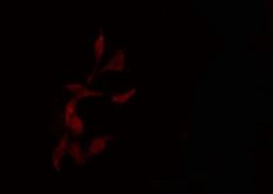
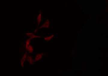
- Immunofluorescent analysis of NCAPG in HepG2 cells. Samples were fixed with paraformaldehyde, permeabilized with 0.1% Triton X-100, blocked with 10% serum (45 min at 25°C) incubated with NCAPG polyclonal antibody (Product # PA5-101540) using a dilution of 1:200 (1 hr, 37°C), and followed by goat anti-rabbit IgG Alexa Fluor 594 at a dilution of 1:600.
- Submitted by
- Invitrogen Antibodies (provider)
- Main image
- Experimental details
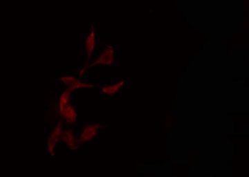
- Immunofluorescent analysis of NCAPG in HepG2 cells. Samples were fixed with paraformaldehyde, permeabilized with 0.1% Triton X-100, blocked with 10% serum (45 min at 25°C) incubated with NCAPG polyclonal antibody (Product # PA5-101540) using a dilution of 1:200 (1 hr, 37°C), and followed by goat anti-rabbit IgG Alexa Fluor 594 at a dilution of 1:600.
Supportive validation
- Submitted by
- Invitrogen Antibodies (provider)
- Main image

- Experimental details
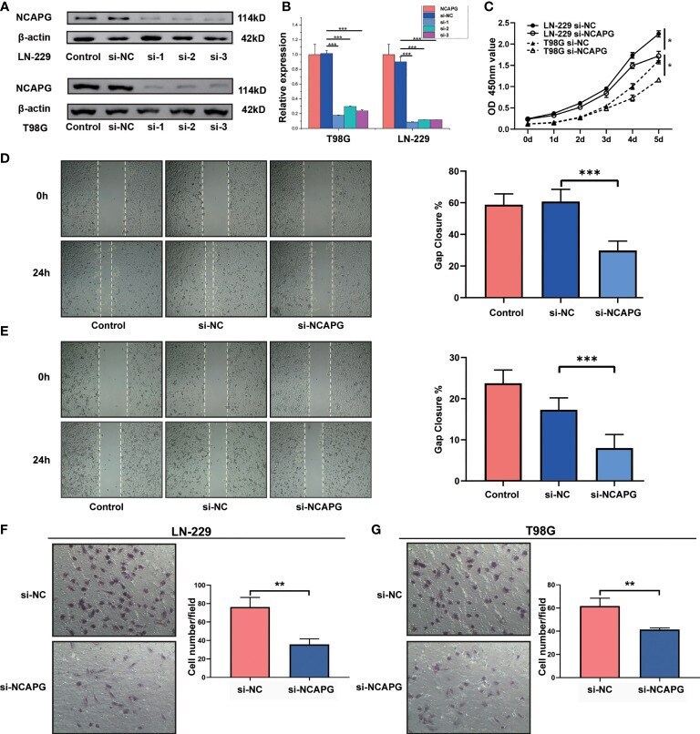
- Figure 4 NCAPG promotes the proliferation, migration, and differentiation of glioma. (A) NCAPG expression in LN-229 and T98G cell lines were analyzed using western blot after transfection with si-NC, si-1, si-2, and si-3. (B) Quantitative analysis of the expression of NCAPG in LN-229 and T98G cell lines by western blot and RT-qPCR. (C) Cell viability of LN-229 and T98G cell lines at different time points after incubation, analyzed by CCK-8. (D, E) Cell scratch test was used to detect the healing rates of LN-229 and T98G cell lines. (F, G) Transwell invasion assay was used to detect the invasion ability of both LN-229 and T98G cell lines. (*P < 0.05, **P < 0.01, ***P < 0.001).
- Submitted by
- Invitrogen Antibodies (provider)
- Main image
- Experimental details
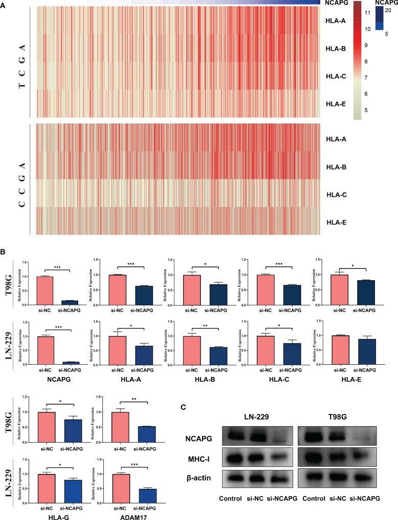
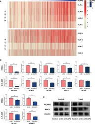
- Figure 7 NCAPG affects the expression of major histocompatibility complex-I (MHC-I) in glioma. (A) Heatmap of the relationship between NCAPG expression and MHC-I in the TCGA and CGGA datasets. (B) The expression of NCAPG in relation to different MHC-I molecules and ADAM17 with RT-qPCR assay. (C) The relationship between NCAPG and MHC-I molecule expression using western blot analysis. (*P < 0.05, **P < 0.01, ***P < 0.001).