Explore
 Validate
 Learn
Learn Western blot
 Other assay
Antibody data
- Antibody Data
- Antigen structure
- References [1]
- Comments [0]
- Validations
- Other assay [1]
Submit
Validation data
Reference
Comment
Report error
- Product number
- PA5-31419 - Provider product page

- Provider
- Invitrogen Antibodies
- Product name
- Fbxo16 Polyclonal Antibody
- Antibody type
- Polyclonal
- Antigen
- Recombinant full-length protein
- Description
- Recommended positive controls: mouse heart. Store product as a concentrated solution. Centrifuge briefly prior to opening the vial.
- Reactivity
- Human, Mouse
- Host
- Rabbit
- Isotype
- IgG
- Vial size
- 100 μL
- Concentration
- 0.61 mg/mL
- Storage
- Store at 4°C short term. For long term storage, store at -20°C, avoiding freeze/thaw cycles.
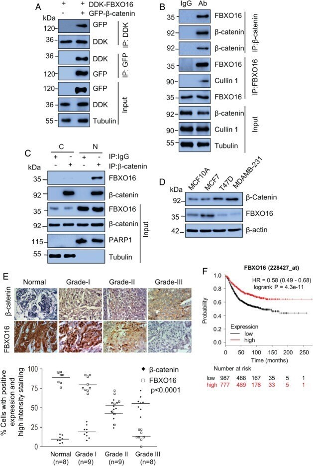
Submitted references F-box protein FBXO16 functions as a tumor suppressor by attenuating nuclear β-catenin function.
Paul D, Islam S, Manne RK, Dinesh US, Malonia SK, Maity B, Boppana R, Rapole S, Shetty PK, Santra MK
The Journal of pathology 2019 Jul;248(3):266-279
The Journal of pathology 2019 Jul;248(3):266-279
No comments: Submit comment
Supportive validation
- Submitted by
- Invitrogen Antibodies (provider)
- Main image
- Experimental details
- Figure 1 FBXO16 interacts with beta-catenin. (A) MCF7 cells coexpressing DDK-FBXO16, either with vector control or GFP-beta-catenin for 40 h, were then incubated with 5 mu m MG132 for 6 h. Whole-cell lysates were immunoprecipitated with the indicated antibodies. Immunoprecipitates and input lysates were separated on SDS-PAGE and immunoblotted for the indicated proteins. Tubulin was used as a loading control throughout our study ( n = 3). (B) Whole-cell lysates of MCF7 cells, pretreated with 5 mu m MG132 for 6 h before harvesting, were immunoprecipitated with the indicated antibodies to pull-down endogenous proteins. IgG immunoprecipitates were used as a control. Immunoprecipitates and input lysates were separated on SDS-PAGE and immunoblotted for the indicated proteins ( n = 2). (C) Nuclear and cytoplasmic fractions were immunoprecipitated with either anti-beta-catenin antibody or IgG. Immunoprecipitates and input lysates were separated on SDS-PAGE and immunoblotted for the indicated proteins ( n = 2). Cells were grown in the presence of 5 mu m MG132 for 6 h before harvesting. (D) Whole-cell protein extracts of MCF10A, MCF7, T47D, and MDA-MB-231 cells were immunoblotted for beta-catenin, FBXO16, and tubulin ( n = 3). beta-actin was used as a loading control. (E) (Top) Expression levels of beta-catenin (top row) and FBXO16 (bottom) in samples of normal and different grades of breast cancer. Tissues were immunostained as described in the 'Materials and methods' section. Lower