Explore
 Validate
 Learn
Learn Western blot
 Immunocytochemistry
Antibody data
- Antibody Data
- Antigen structure
- References [8]
- Comments [0]
- Validations
- Immunocytochemistry [2]
- Immunohistochemistry [2]
- Other assay [6]
Submit
Validation data
Reference
Comment
Report error
- Product number
- PA5-22142 - Provider product page

- Provider
- Invitrogen Antibodies
- Product name
- FIS1 Polyclonal Antibody
- Antibody type
- Polyclonal
- Antigen
- Recombinant full-length protein
- Description
- Recommended positive controls: 293T, A431, HeLa, HepG2, Raji, Mouse brain, mouse liver, rat brain. Predicted reactivity: Mouse (97%), Rat (97%), Rhesus Monkey (100%), Bovine (97%).
- Reactivity
- Human, Mouse, Rat
- Host
- Rabbit
- Isotype
- IgG
- Vial size
- 100 μL
- Concentration
- 1.4 mg/mL
- Storage
- Store at 4°C short term. For long term storage, store at -20°C, avoiding freeze/thaw cycles.
Submitted references Characterisation of cold-induced mitochondrial fission in porcine aortic endothelial cells.
All-Trans Retinoic Acid Increases DRP1 Levels and Promotes Mitochondrial Fission.
Changes in Drp1 Function and Mitochondrial Morphology Are Associated with the α-Synuclein Pathology in a Transgenic Mouse Model of Parkinson's Disease.
Deletion of Mitochondrial Uncoupling Protein 2 Exacerbates Mitochondrial Damage in Mice Subjected to Cerebral Ischemia and Reperfusion Injury under both Normo- and Hyperglycemic Conditions.
Inhibition of chymotrypsin-like activity of the proteasome by ixazomib prevents mitochondrial dysfunction during myocardial ischemia.
Azoramide protects iPSC-derived dopaminergic neurons with PLA2G6 D331Y mutation through restoring ER function and CREB signaling.
Infection-Induced Peroxisome Biogenesis Is a Metabolic Strategy for Herpesvirus Replication.
Ubiquitin-mediated regulation of the E3 ligase GP78 by MGRN1 in trans affects mitochondrial homeostasis.
Quiring L, Walter B, Lohaus N, Schwan D, Rech A, Dlugos A, Rauen U
Molecular medicine (Cambridge, Mass.) 2022 Jan 31;28(1):13
Molecular medicine (Cambridge, Mass.) 2022 Jan 31;28(1):13
All-Trans Retinoic Acid Increases DRP1 Levels and Promotes Mitochondrial Fission.
Chidipi B, Shah SI, Reiser M, Kanithi M, Garces A, Cha BJ, Ullah G, Noujaim SF
Cells 2021 May 14;10(5)
Cells 2021 May 14;10(5)
Changes in Drp1 Function and Mitochondrial Morphology Are Associated with the α-Synuclein Pathology in a Transgenic Mouse Model of Parkinson's Disease.
Portz P, Lee MK
Cells 2021 Apr 13;10(4)
Cells 2021 Apr 13;10(4)
Deletion of Mitochondrial Uncoupling Protein 2 Exacerbates Mitochondrial Damage in Mice Subjected to Cerebral Ischemia and Reperfusion Injury under both Normo- and Hyperglycemic Conditions.
He M, Ma Y, Wang R, Zhang J, Jing L, Li PA
International journal of biological sciences 2020;16(15):2788-2802
International journal of biological sciences 2020;16(15):2788-2802
Inhibition of chymotrypsin-like activity of the proteasome by ixazomib prevents mitochondrial dysfunction during myocardial ischemia.
Sánchez G, Chalmers S, Ahumada X, Montecinos L, Olmedo I, Eisner V, Riveros A, Kogan MJ, Lavandero S, Pedrozo Z, Donoso P
PloS one 2020;15(5):e0233591
PloS one 2020;15(5):e0233591
Azoramide protects iPSC-derived dopaminergic neurons with PLA2G6 D331Y mutation through restoring ER function and CREB signaling.
Ke M, Chong CM, Zeng H, Huang M, Huang Z, Zhang K, Cen X, Lu JH, Yao X, Qin D, Su H
Cell death & disease 2020 Feb 18;11(2):130
Cell death & disease 2020 Feb 18;11(2):130
Infection-Induced Peroxisome Biogenesis Is a Metabolic Strategy for Herpesvirus Replication.
Jean Beltran PM, Cook KC, Hashimoto Y, Galitzine C, Murray LA, Vitek O, Cristea IM
Cell host & microbe 2018 Oct 10;24(4):526-541.e7
Cell host & microbe 2018 Oct 10;24(4):526-541.e7
Ubiquitin-mediated regulation of the E3 ligase GP78 by MGRN1 in trans affects mitochondrial homeostasis.
Mukherjee R, Chakrabarti O
Journal of cell science 2016 Feb 15;129(4):757-73
Journal of cell science 2016 Feb 15;129(4):757-73
No comments: Submit comment
Supportive validation
- Submitted by
- Invitrogen Antibodies (provider)
- Main image

- Experimental details
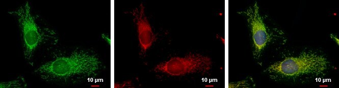
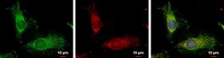
- FIS1 Polyclonal Antibody detects FIS1 protein at mitochondria by immunofluorescent analysis. Sample: HeLa cells were fixed in 2% paraformaldehyde/culture medium at 37oC for 30 min. Green: FIS1 protein stained by FIS1 Polyclonal Antibody (Product # PA5-22142) diluted at 1:2,000. Red: MitoTrackerR Red CMXRos, a mitochondria tracker. Blue: Hoechst 33342 staining. Scale bar = 10 µm.
- Submitted by
- Invitrogen Antibodies (provider)
- Main image
- Experimental details
- FIS1 Polyclonal Antibody detects FIS1 protein at mitochondria by immunofluorescent analysis. Sample: HeLa cells were fixed in 2% paraformaldehyde/culture medium at 37oC for 30 min. Green: FIS1 protein stained by FIS1 Polyclonal Antibody (Product # PA5-22142) diluted at 1:2,000. Red: MitoTrackerR Red CMXRos, a mitochondria tracker. Blue: Hoechst 33342 staining. Scale bar = 10 µm.

Supportive validation
- Submitted by
- Invitrogen Antibodies (provider)
- Main image
- Experimental details
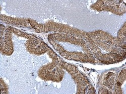
- TTC11 antibody detects TTC11 protein at mitochondria on mouse prostate by immunohistochemical analysis. Sample: Paraffin-embedded mouse prostate. TTC11 antibody (Product # PA5-22142) dilution: 1:500. Antigen Retrieval: EDTA based buffer, pH 8.0, 15 min.

- Submitted by
- Invitrogen Antibodies (provider)
- Main image
- Experimental details
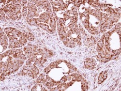
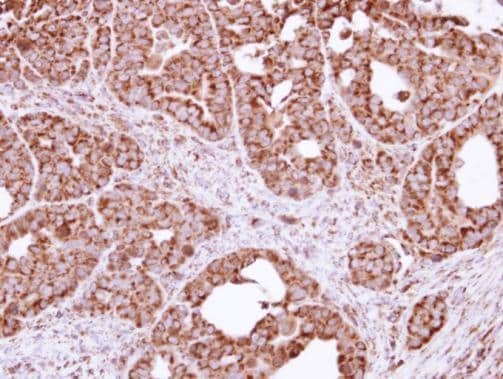
- Immunohistochemical analysis of paraffin-embedded NCIN87 xenograft, using FIS1 (Product # PA5-22142) antibody at 1:100 dilution. Antigen Retrieval: EDTA based buffer, pH 8.0, 15 min.
Supportive validation
- Submitted by
- Invitrogen Antibodies (provider)
- Main image
- Experimental details
- NULL
- Submitted by
- Invitrogen Antibodies (provider)
- Main image
- Experimental details
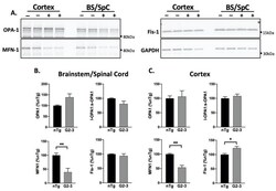
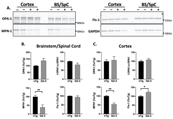
- Figure 4 Decreased MFN1 levels with A53T mutant human alphaS expression in vivo. ( A ) Western blot of mitochondrial dynamics proteins in the total brain lysate of nTg (-) and Tg (+) animals. ( B , C ) A quantification of immunoblots shown in ( A ) normalized to GAPDH, show a decrease in MFN1 levels in the BS/SpC and the Ctx ( p = 0.0074, p = 0018, respectively) of the TgA53TG2-3(G2-3) samples compared to the nTg samples. In the G2-3 cortex, Fis-1 levels were also increased ( p = 0.0145), per the unpaired t -tests with Welch''s correction ( n = 5 per group, mean +- SEM). (* p < 0.05, ** p < 0.01).
- Submitted by
- Invitrogen Antibodies (provider)
- Main image
- Experimental details
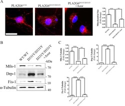
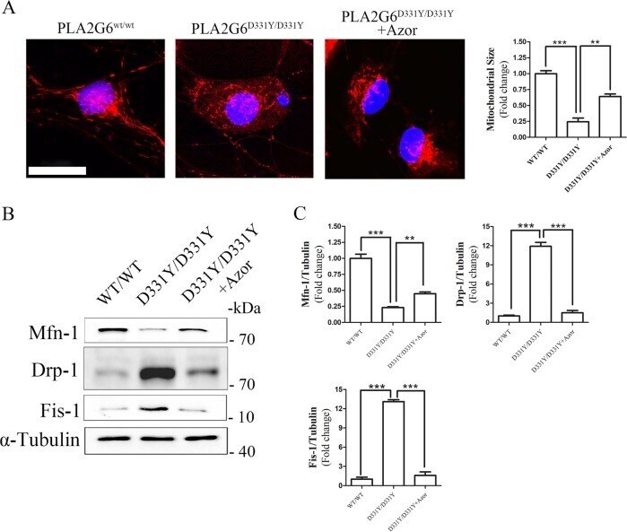
- Fig. 4 Azoramide prevents mitochondrial fragmentation in PLA2G6 D331Y/D331Y DA neurons. a PLA2G6 D331Y/D331Y DA neurons were treated with 10 muM azoramide from day 15 to 20. Representative images and quantification of mitochondria in PLA2G6 WT/WT , PLA2G6 D331Y/D331Y and azoramide-treated PLA2G6 D331Y/D331Y DA neurons after MitoTracker Red staining. b , c Expression of mitochondrial fission and fusion proteins was determined and quantified by western blotting. alpha-Tubulin was the internal control. Data are represented as mean +- SEM. ** p < 0.01, *** p < 0.005. Scale bar: 20 um.
- Submitted by
- Invitrogen Antibodies (provider)
- Main image
- Experimental details
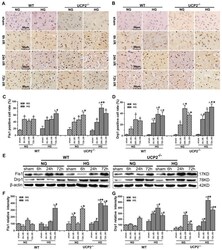
- Figure 5 UCP2 deletion increased the levels of mitochondrial fission-related proteins in hyperglycemic mice after ischemia/reperfusion. Detections of mitochondrial fission-related proteins by immunohistochemistry and Western blotting (n=6, per group). (A, B) Representative photomicrographs for Fis1 and Drp1. Scale bar=50 mum. (C, D) Quantification of Fis1 and Drp1 immunointensity. E: Representative Western blots for Fis1 and Drp1. (F, G) Semi-quantification of Fis1 and Drp1 protein bands. Data are shown as mean +- SD. For values in F and G, the values in WT+NG sham group were converted to 100 and percent changes were presented for other groups relative to the WT+NG sham. Delta p
- Submitted by
- Invitrogen Antibodies (provider)
- Main image
- Experimental details
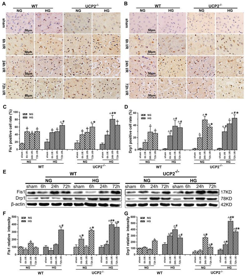
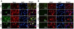
- Figure 8 Mitochondrial fusion-related proteins (Opa1 and Mfn2) and fission-related proteins (Drp1, Fis1) co-localized with neurons in UCP2 -/- mice following ischemic stroke. Double immunostaining of Drp1, Fis1, Opa1 and Mfn2 with GFAP (astrocyte marker) and NeuN (neuron marker) were performed in UCP2 -/- mice brain sections 1 day after reperfusion (n=6, each group). Scale bar =25 mum.
- Submitted by
- Invitrogen Antibodies (provider)
- Main image

- Experimental details
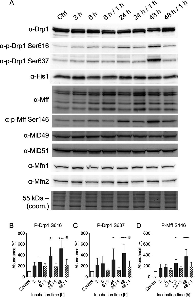
- Fig. 6 Qualitative and quantitative analysis of proteins involved in mitochondrial fission and outer membrane fusion. Protein lysates of porcine aortic endothelial cells incubated for 3 h, 6 h, 24 h or 48 h at 4 degC and rewarmed for 1 h at 37 degC were analysed by western blot ( A ) for protein levels of Drp1 and phosphorylation of Drp1 (activating site p-Drp1-S616 and inactivating site p-Drp1-S637) and Fis1. Additionally Mff, p-Mff Ser146, MiD49, MiD51, Mfn1 and Mfn2 were targeted. Coomassie staining was used as loading control (coom.). Representative figures of n = 4 experiments. The band intensity was quantified using the BioVision software and normalized to the control ( B - D ). Rewarming conditions are shown hatched. Means +- SD of n = 4 experiments. * Significantly different to control (p