Explore
 Validate
 Learn
Learn Western blot
 Immunocytochemistry
Antibody data
- Antibody Data
- Antigen structure
- References [14]
- Comments [0]
- Validations
- Immunocytochemistry [1]
Submit
Validation data
Reference
Comment
Report error
- Product number
- HPA017430 - Provider product page

- Provider
- Atlas Antibodies
- Proper citation
- Atlas Antibodies Cat#HPA017430, RRID:AB_1848608
- Product name
- Anti-FIS1
- Antibody type
- Polyclonal
- Description
- Polyclonal Antibody against Human FIS1, Gene description: fission 1 (mitochondrial outer membrane) homolog (S. cerevisiae), Alternative Gene Names: CGI-135, Fis1, H_NH0132A01.6, TTC11, Validated applications: WB, IHC, ICC, Uniprot ID: Q9Y3D6, Storage: Store at +4°C for short term storage. Long time storage is recommended at -20°C.
- Reactivity
- Human, Mouse, Rat
- Host
- Rabbit
- Conjugate
- Unconjugated
- Isotype
- IgG
- Vial size
- 100 µl
- Concentration
- 0.2 mg/ml
- Storage
- Store at +4°C for short term storage. Long time storage is recommended at -20°C.
- Handling
- The antibody solution should be gently mixed before use.
Submitted references Melatonin Improves Skeletal Muscle Structure and Oxidative Phenotype by Regulating Mitochondrial Dynamics and Autophagy in Zücker Diabetic Fatty Rat
Flow pattern–dependent mitochondrial dynamics regulates the metabolic profile and inflammatory state of endothelial cells
Acute resistance exercise training does not augment mitochondrial remodelling in master athletes or untrained older adults.
NIX initiates mitochondrial fragmentation via DRP1 to drive epidermal differentiation
MIEF1/2 orchestrate mitochondrial dynamics through direct engagement with both the fission and fusion machineries.
Short-term step reduction reduces citrate synthase activity without altering skeletal muscle markers of oxidative metabolism or insulin-mediated signaling in young males.
The vitamin D receptor regulates mitochondrial function in C2C12 myoblasts
Gas7 knockout affects PINK1 expression and mitochondrial dynamics in mouse cortical neurons
Actin chromobody imaging reveals sub-organellar actin dynamics.
Mitochondrial fragmentation affects neither the sensitivity to TNFα-induced apoptosis of Brucella-infected cells nor the intracellular replication of the bacteria
Impaired muscle relaxation and mitochondrial fission associated with genetic ablation of cytoplasmic actin isoforms
Skeletal Muscle Fibre-Specific Knockout of p53 Does Not Reduce Mitochondrial Content or Enzyme Activity
A ketogenic diet accelerates neurodegeneration in mice with induced mitochondrial DNA toxicity in the forebrain
Different molecular mechanisms involved in spontaneous and oxidative stress-induced mitochondrial fragmentation in tripeptidyl peptidase-1 (TPP-1)-deficient fibroblasts
Salagre D, Raya Álvarez E, Cendan C, Aouichat S, Agil A
Antioxidants 2023;12(8):1499
Antioxidants 2023;12(8):1499
Flow pattern–dependent mitochondrial dynamics regulates the metabolic profile and inflammatory state of endothelial cells
Hong S, Shin J, Choi S, Powers J, Meister B, Sayoc J, Son J, Tierney R, Recchia F, Brown M, Yang X, Park J
JCI Insight 2022;7(18)
JCI Insight 2022;7(18)
Acute resistance exercise training does not augment mitochondrial remodelling in master athletes or untrained older adults.
Marshall RN, McKendry J, Smeuninx B, Seabright AP, Morgan PT, Greig C, Breen L
Frontiers in physiology 2022;13:1097988
Frontiers in physiology 2022;13:1097988
NIX initiates mitochondrial fragmentation via DRP1 to drive epidermal differentiation
Simpson C, Tokito M, Uppala R, Sarkar M, Gudjonsson J, Holzbaur E
Cell Reports 2021;34(5):108689
Cell Reports 2021;34(5):108689
MIEF1/2 orchestrate mitochondrial dynamics through direct engagement with both the fission and fusion machineries.
Yu R, Liu T, Jin SB, Ankarcrona M, Lendahl U, Nistér M, Zhao J
BMC biology 2021 Oct 21;19(1):229
BMC biology 2021 Oct 21;19(1):229
Short-term step reduction reduces citrate synthase activity without altering skeletal muscle markers of oxidative metabolism or insulin-mediated signaling in young males.
Edwards SJ, Shad BJ, Marshall RN, Morgan PT, Wallis GA, Breen L
Journal of applied physiology (Bethesda, Md. : 1985) 2021 Dec 1;131(6):1653-1662
Journal of applied physiology (Bethesda, Md. : 1985) 2021 Dec 1;131(6):1653-1662
The vitamin D receptor regulates mitochondrial function in C2C12 myoblasts
Ashcroft S, Bass J, Kazi A, Atherton P, Philp A
American Journal of Physiology-Cell Physiology 2020;318(3):C536-C541
American Journal of Physiology-Cell Physiology 2020;318(3):C536-C541
Gas7 knockout affects PINK1 expression and mitochondrial dynamics in mouse cortical neurons
Bhupana J, Huang B, Liou G, Calkins M, Lin‐Chao S
FASEB BioAdvances 2020;2(3):166-181
FASEB BioAdvances 2020;2(3):166-181
Actin chromobody imaging reveals sub-organellar actin dynamics.
Schiavon CR, Zhang T, Zhao B, Moore AS, Wales P, Andrade LR, Wu M, Sung TC, Dayn Y, Feng JW, Quintero OA, Shadel GS, Grosse R, Manor U
Nature methods 2020 Sep;17(9):917-921
Nature methods 2020 Sep;17(9):917-921
Mitochondrial fragmentation affects neither the sensitivity to TNFα-induced apoptosis of Brucella-infected cells nor the intracellular replication of the bacteria
Lobet E, Willemart K, Ninane N, Demazy C, Sedzicki J, Lelubre C, De Bolle X, Renard P, Raes M, Dehio C, Letesson J, Arnould T
Scientific Reports 2018;8(1)
Scientific Reports 2018;8(1)
Impaired muscle relaxation and mitochondrial fission associated with genetic ablation of cytoplasmic actin isoforms
O'Rourke A, Lindsay A, Tarpey M, Yuen S, McCourt P, Nelson D, Perrin B, Thomas D, Spangenburg E, Lowe D, Ervasti J
The FEBS Journal 2018;285(3):481-500
The FEBS Journal 2018;285(3):481-500
Skeletal Muscle Fibre-Specific Knockout of p53 Does Not Reduce Mitochondrial Content or Enzyme Activity
Stocks B, Dent J, Joanisse S, McCurdy C, Philp A
Frontiers in Physiology 2017;8
Frontiers in Physiology 2017;8
A ketogenic diet accelerates neurodegeneration in mice with induced mitochondrial DNA toxicity in the forebrain
Lauritzen K, Hasan-Olive M, Regnell C, Kleppa L, Scheibye-Knudsen M, Gjedde A, Klungland A, Bohr V, Storm-Mathisen J, Bergersen L
Neurobiology of Aging 2016;48
Neurobiology of Aging 2016;48
Different molecular mechanisms involved in spontaneous and oxidative stress-induced mitochondrial fragmentation in tripeptidyl peptidase-1 (TPP-1)-deficient fibroblasts
Van Beersel G, Tihon E, Demine S, Hamer I, Jadot M, Arnould T
Bioscience Reports 2013;33(2)
Bioscience Reports 2013;33(2)
No comments: Submit comment
Supportive validation
- Submitted by
- Atlas Antibodies (provider)
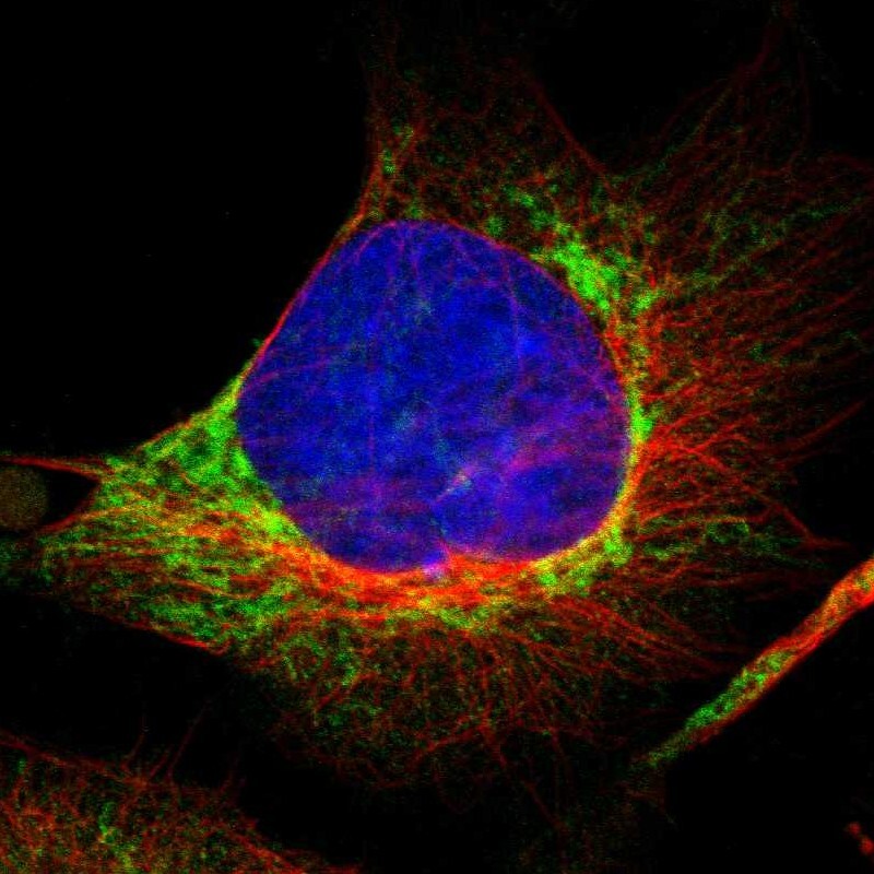
- Main image

- Experimental details
- Immunofluorescent staining of human cell line U-251 MG shows localization to mitochondria.
- Sample type
- Human