Explore
 Validate
 Learn
Learn Western blot
Antibody data
- Antibody Data
- Antigen structure
- References [1]
- Comments [0]
- Validations
- Western blot [3]
- Immunocytochemistry [1]
Submit
Validation data
Reference
Comment
Report error
- Product number
- GTX103192 - Provider product page

- Provider
- GeneTex
- Proper citation
- GeneTex Cat#GTX103192, RRID:AB_1951628
- Product name
- RanBP1 antibody [N1C3]
- Antibody type
- Polyclonal
- Reactivity
- Human, Mouse
- Host
- Rabbit
Submitted references NMDAR signaling facilitates the IPO5-mediated nuclear import of CPEB3.
Chao HW, Lai YT, Lu YL, Lin CL, Mai W, Huang YS
Nucleic acids research 2012 Sep 1;40(17):8484-98
Nucleic acids research 2012 Sep 1;40(17):8484-98
No comments: Submit comment
Supportive validation
- Submitted by
- GeneTex (provider)
- Main image

- Experimental details
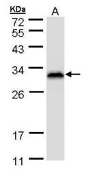
- Sample (50 ug of whole cell lysate) A: Mouse brain 12% SDS PAGE GTX103192 diluted at 1:1000

- Submitted by
- GeneTex (provider)
- Main image
- Experimental details
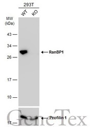
- Sample (30 ug of whole cell lysate)A: HeLa S3 (GTX14654)12% SDS PAGEGTX103192 diluted at 1:1000

- Submitted by
- GeneTex (provider)
- Main image
- Experimental details
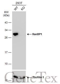
- Wild-type (WT) and RanBP1 knockout (KO) 293T cell extracts (30 ?g) were separated by 12% SDS-PAGE, and the membrane was blotted with RanBP1 antibody [N1C3] (GTX103192) diluted at 1:2000. The HRP-conjugated anti-rabbit IgG antibody (GTX213110-01) was used to detect the primary antibody.
Supportive validation
- Submitted by
- GeneTex (provider)
- Main image

- Experimental details
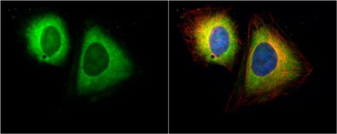
- Ran BP1 antibody [N1C3] detects Ran BP1 protein at cytoplasm by immunofluorescent analysis.Sample: HeLa cells were fixed in 4% paraformaldehyde at RT for 15 min.Green: Ran BP1 protein stained by Ran BP1 antibody [N1C3] (GTX103192) diluted at 1:500.Red: alpha Tubulin, a cytoskeleton marker, stained by alpha Tubulin antibody [B-5-1-2] (GTX11304) diluted at 1:10000.Blue: Hoechst 33342 staining.