 Explore
 Validate
 Learn
Learn Western blot
Antibody data
- Antibody Data
- Antigen structure
- References [0]
- Comments [0]
- Validations
- Western blot [1]
- Immunocytochemistry [1]
- Immunohistochemistry [1]
Submit
Validation data
Reference
Comment
Report error
- Product number
- TA328650 - Provider product page

- Provider
- OriGene
- Product name
- Rabbit Polyclonal Anti-Nogo Receptor
- Antibody type
- Polyclonal
- Description
- Rabbit Polyclonal Anti-Nogo Receptor
- Host
- Rabbit
- Conjugate
- Unconjugated
- Epitope
- RTN4R
- Antibody clone number
- NULL
- Vial size
- 200 µl
- Concentration
- NULL
No comments: Submit comment
Supportive validation
- Submitted by
- OriGene (provider)
- Main image
- Experimental details
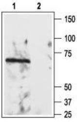
- Western blot analysis of rat brain lysate: 1. Anti-Nogo Receptor (extracellular) antibody, (1:200). 2. Anti-Nogo Receptor (extracellular) antibody, preincubated with the control peptide antigen.
- Validation comment
- WB

Supportive validation
- Submitted by
- OriGene (provider)
- Main image
- Experimental details
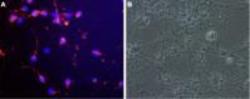
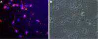
- Expression of Nogo receptor in rat cerebellar granule. Immunocytochemical staining of live cultured rat cerebellar granule. A. Cells were stained with Anti-Nogo Receptor (extracellular) antibody followed by goat-anti-rabbit AlexaFluor-555 secondary antibody (red). Nuclei were visualized with the cell permeable dye Hoechst 33342 (blue). B. Live view of the same field as in A.
- Validation comment
- IF
Supportive validation
- Submitted by
- OriGene (provider)
- Main image
- Experimental details
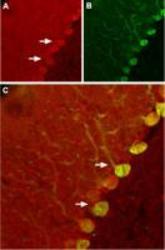
- Expression of Nogo receptor in rat cerebellum. Immunohistochemical staining of rat cerebellum using Anti-Nogo Receptor (extracellular) antibody.A. Nogo receptor (red) appears in Purkinje cells (arrows). B. Staining of Purkinje nerve cells with mouse anti-calbindin D28K (a calcium binding protein, green). C. Confocal merge of Nogo receptor and calbindin D28K demonstrates some co-localization of these proteins.
- Validation comment
- IHC
