Explore
 Validate
 Learn
Learn Western blot
Antibody data
- Antibody Data
- Antigen structure
- References [0]
- Comments [0]
- Validations
- Western blot [1]
- Immunocytochemistry [1]
- Immunohistochemistry [2]
- Flow cytometry [1]
Submit
Validation data
Reference
Comment
Report error
- Product number
- ANT-008-25UL - Provider product page

- Provider
- Invitrogen Antibodies
- Product name
- Nogo Receptor (extracellular) Polyclonal Antibody
- Antibody type
- Polyclonal
- Antigen
- Other
- Reactivity
- Human, Mouse, Rat
- Host
- Rabbit
- Isotype
- IgG
- Vial size
- 25 µL
- Concentration
- 0.8 mg/mL
- Storage
- -20° C, Avoid Freeze/Thaw Cycles
No comments: Submit comment
Supportive validation
- Submitted by
- Invitrogen Antibodies (provider)
- Main image

- Experimental details
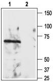
- Western blot analysisof rat brain lysate: - 1. Anti-Nogo Receptor (extracellular) Antibody (#ANT-008), (1:200). 2. Anti-Nogo Receptor (extracellular) Antibody , preincubated with Nogo Receptor (extracellular) Blocking Peptide (#BLP-NT008).
Supportive validation
- Submitted by
- Invitrogen Antibodies (provider)
- Main image
- Experimental details
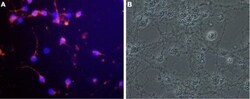
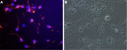
- Expression of Nogo receptor in rat cerebellar granule - Cell surface detection of Nogo receptor in live cultured rat cerebellar granule. A. Cells were stained with Anti-Nogo Receptor (extracellular) Antibody (#ANT-008) followed by goat- Anti-rabbit AlexaFluor-555 secondary Antibody (red). Nuclei were visualized with the cell permeable dye Hoechst 33342 (blue). B.Live view of the same field as in A.
Supportive validation
- Submitted by
- Invitrogen Antibodies (provider)
- Main image
- Experimental details
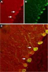
- Expression of Nogo receptor in rat cerebellum - Immunohistochemical staining of rat cerebellum using Anti-Nogo Receptor (extracellular) Antibody (#ANT-008). A. NgR (red) appears in Purkinje cells (arrows). B. Staining of Purkinje nerve cells with mouse Anti-calbindin D28K (a calcium binding protein, green). C. Confocal merge of NgR and calbindin D28K demonstrates some co-localization of these proteins.
- Submitted by
- Invitrogen Antibodies (provider)
- Main image

- Experimental details
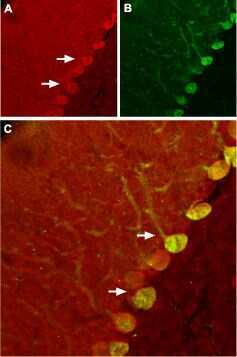
- Expression of Nogo receptor in rat cerebellum - Immunohistochemical staining of rat cerebellum using Anti-Nogo Receptor (extracellular) Antibody (#ANT-008). A. NgR (red) appears in Purkinje cells (arrows). B. Staining of Purkinje nerve cells with mouse Anti-calbindin D28K (a calcium binding protein, green). C. Confocal merge of NgR and calbindin D28K demonstrates some co-localization of these proteins.

Supportive validation
- Submitted by
- Invitrogen Antibodies (provider)
- Main image
- Experimental details
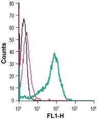
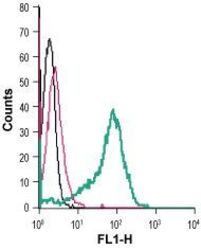
- Cell surface detection of Nogo Receptor in live intact human THP-1 monocytic leukemia cells: - (black line) cells. (red) Cells + goat- Anti-rabbit-FITC. (green) Cells + Anti-Nogo Receptor (extracellular) Antibody (#ANT-008), (2.5 µg) + goat- Anti-rabbit-FITC.