Explore
 Validate
 Learn
Learn Western blot
 Immunocytochemistry
Antibody data
- Antibody Data
- Antigen structure
- References [2]
- Comments [0]
- Validations
- Immunocytochemistry [2]
- Immunohistochemistry [6]
- Other assay [1]
Submit
Validation data
Reference
Comment
Report error
- Product number
- PA5-31231 - Provider product page

- Provider
- Invitrogen Antibodies
- Product name
- RBPMS Polyclonal Antibody
- Antibody type
- Polyclonal
- Antigen
- Recombinant full-length protein
- Description
- Recommended positive controls: A549, mouse heart. Predicted reactivity: Mouse (99%), Rat (99%), Bovine (98%). Store product as a concentrated solution. Centrifuge briefly prior to opening the vial.
- Reactivity
- Human, Mouse, Rat
- Host
- Rabbit
- Isotype
- IgG
- Vial size
- 100 μL
- Concentration
- 1.34 mg/mL
- Storage
- Store at 4°C short term. For long term storage, store at -20°C, avoiding freeze/thaw cycles.
Submitted references Genetic inhibition of collapsin response mediator protein-2 phosphorylation ameliorates retinal ganglion cell death in normal-tension glaucoma models.
Genetic inhibition of CRMP2 phosphorylation at serine 522 promotes axonal regeneration after optic nerve injury.
Brahma MM, Takahashi K, Namekata K, Harada T, Goshima Y, Ohshima T
Genes to cells : devoted to molecular & cellular mechanisms 2022 Aug;27(8):526-536
Genes to cells : devoted to molecular & cellular mechanisms 2022 Aug;27(8):526-536
Genetic inhibition of CRMP2 phosphorylation at serine 522 promotes axonal regeneration after optic nerve injury.
Kondo S, Takahashi K, Kinoshita Y, Nagai J, Wakatsuki S, Araki T, Goshima Y, Ohshima T
Scientific reports 2019 May 10;9(1):7188
Scientific reports 2019 May 10;9(1):7188
No comments: Submit comment
Supportive validation
- Submitted by
- Invitrogen Antibodies (provider)
- Main image

- Experimental details
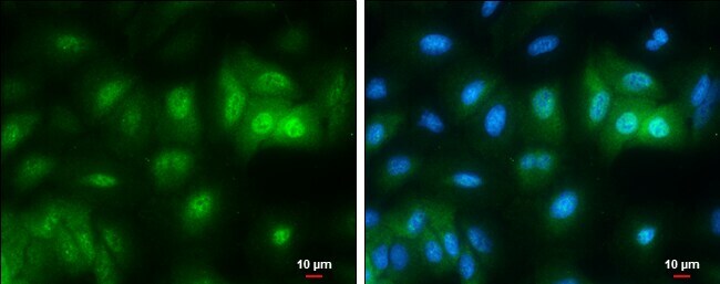
- Immunocytochemistry-Immunofluorescence analysis of RBPMS was performed in A549 cells fixed in 4% paraformaldehyde at RT for 15 min. Green: RBPMS Polyclonal Antibody (Product # PA5-31231) diluted at 1:500. Blue: Hoechst 33342 staining. Scale bar = 10 µm.
- Submitted by
- Invitrogen Antibodies (provider)
- Main image
- Experimental details
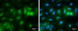
- Immunocytochemistry-Immunofluorescence analysis of RBPMS was performed in A549 cells fixed in 4% paraformaldehyde at RT for 15 min. Green: RBPMS Polyclonal Antibody (Product # PA5-31231) diluted at 1:500. Blue: Hoechst 33342 staining. Scale bar = 10 µm.

Supportive validation
- Submitted by
- Invitrogen Antibodies (provider)
- Main image

- Experimental details
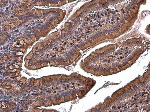
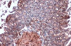
- Immunohistochemistry (Paraffin) analysis of RBPMS was performed in paraffin-embedded rat duodenum tissue using RBPMS Polyclonal Antibody (Product # PA5-31231) at a dilution of 1:500.
- Submitted by
- Invitrogen Antibodies (provider)
- Main image
- Experimental details
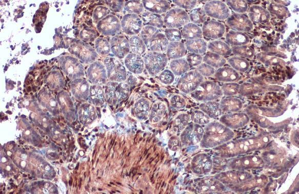
- Immunohistochemistry (Paraffin) analysis of RBPMS was performed in paraffin-embedded mouse intestine tissue using RBPMS Polyclonal Antibody (Product # PA5-31231) at a dilution of 1:2000. Antigen Retrieval: Citrate buffer, pH 6.0, 15 min.
- Submitted by
- Invitrogen Antibodies (provider)
- Main image

- Experimental details
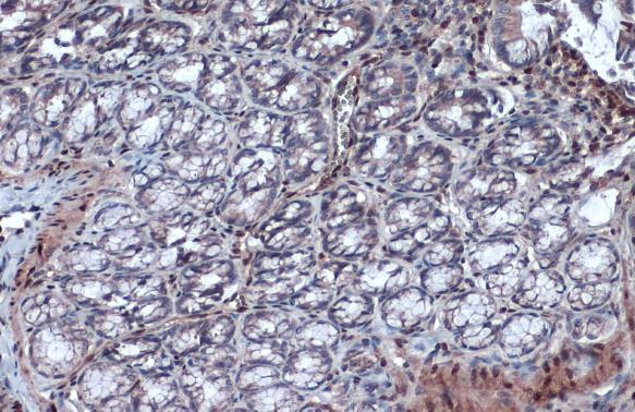
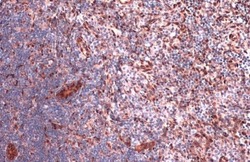
- Immunohistochemistry (Paraffin) analysis of RBPMS was performed in paraffin-embedded rat colon tissue using RBPMS Polyclonal Antibody (Product # PA5-31231) at a dilution of 1:500. Antigen Retrieval: Citrate buffer, pH 6.0, 15 min.
- Submitted by
- Invitrogen Antibodies (provider)
- Main image

- Experimental details
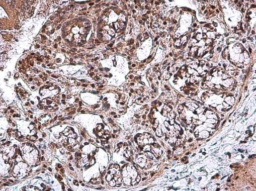
- Immunohistochemistry (Paraffin) analysis of RBPMS was performed in paraffin-embedded rat colon tissue using RBPMS Polyclonal Antibody (Product # PA5-31231) at a dilution of 1:500.
- Submitted by
- Invitrogen Antibodies (provider)
- Main image
- Experimental details
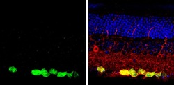
- Immunohistochemistry (Paraffin) analysis of RBPMS was performed in paraffin-embedded mouse retina tissue. Green: RBPMS stained by RBPMS Polyclonal Antibody (Product # PA5-31231) at a dilution of 1:250. Red: beta Tubulin 3/ Tuj1, a marker, stained by beta Tubulin 3/ Tuj1 antibody. Blue: Fluoroshield with DAPI. Antigen Retrieval: Citrate buffer, pH 6.0, 15 min.

- Submitted by
- Invitrogen Antibodies (provider)
- Main image
- Experimental details
- Immunohistochemistry (Paraffin) analysis of RBPMS was performed in paraffin-embedded rat lymph node tissue using RBPMS Polyclonal Antibody (Product # PA5-31231) at a dilution of 1:2000. Antigen Retrieval: Citrate buffer, pH 6.0, 15 min.

Supportive validation
- Submitted by
- Invitrogen Antibodies (provider)
- Main image
- Experimental details
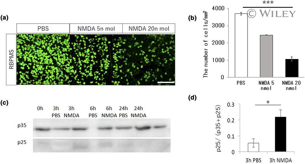
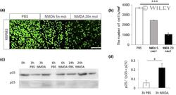
- 1 FIGURE Production of p25 after N-methyl-D-aspartate (NMDA) injection. (a, b) Dose-dependent decrease in the number of retinal ganglion cells (RGCs) observed 1 week after the injection of phosphate-buffered saline (PBS) or NMDA in 5 or 20 nmol concentrations. The retina samples are stained with anti-RNA-binding protein with multiple splicing, a specific marker for RGCs (Rodriguez et al., 2015). (c) Time course of p25 production within 24 h and amounts of p35 and p25 in retina samples from PBS or 20 nmol NMDA injected in the eye indicated time points by western blot analysis. (d) Quantification of p25/p35+p25 level ( n = 4, *** p < .001; * p < .05)