Explore
 Validate
 Learn
Learn50-9856-42
antibody from Invitrogen Antibodies
Targeting: CLEC7A
CD369, CLECSF12, dectin-1, hDectin-1, SCARE2
 Flow cytometry
Antibody data
- Antibody Data
- Antigen structure
- References [7]
- Comments [0]
- Validations
- Flow cytometry [1]
- Other assay [3]
Submit
Validation data
Reference
Comment
Report error
- Product number
- 50-9856-42 - Provider product page

- Provider
- Invitrogen Antibodies
- Product name
- CD369 (Clec7a, Dectin-1) Monoclonal Antibody (15E2), eFluor™ 660, eBioscience™
- Antibody type
- Monoclonal
- Antigen
- Other
- Description
- Description: This 15E2 monoclonal antibody reacts with human and non-human primate CD369, which is also known as the Dectin-1 or beta-glucan receptor or CLEC7A. CD369 is expressed predominantly on monocytes, macrophages, and dendritic cells. Moreover, this receptor can also be detected on neutrophils and lymphocytes, as well as induced by cytokines such as IL-4 or LPS. In humans, CD369 exists as two isoforms that arise due to alternative splicing. CD369 binds beta-1,3-glucan and plays an important role in activating signal transduction involved in the innate immune response to fungal and mycobacterial infections. Studies have also shown the involvement of CD369 in dendritic cell activation.
- Antibody clone number
- 15E2
- Concentration
- 5 µL/Test
Submitted references High-Yield Human Induced Pluripotent Stem Cell-Derived Monocytes and Macrophages Are Functionally Comparable With Primary Cells.
Isoform localization of Dectin-1 regulates the signaling quality of anti-fungal immunity.
MiR-146a negatively regulates dectin-1-induced inflammatory responses.
Macrophage dectin-1 expression is controlled by leukotriene B4 via a GM-CSF/PU.1 axis.
Dectin-1 is an extracellular pathogen sensor for the induction and processing of IL-1β via a noncanonical caspase-8 inflammasome.
Concomitant activation and antigen uptake via human dectin-1 results in potent antigen-specific CD8+ T cell responses.
Dectin-1 and its role in the recognition of beta-glucans by macrophages.
Cui D, Franz A, Fillon SA, Jannetti L, Isambert T, Fundel-Clemens K, Huber HJ, Viollet C, Ghanem A, Niwa A, Weigle B, Pflanz S
Frontiers in cell and developmental biology 2021;9:656867
Frontiers in cell and developmental biology 2021;9:656867
Isoform localization of Dectin-1 regulates the signaling quality of anti-fungal immunity.
Fischer M, Müller JP, Spies-Weisshart B, Gräfe C, Kurzai O, Hünniger K, Hochhaus A, Scholl S, Schnetzke U
European journal of immunology 2017 May;47(5):848-859
European journal of immunology 2017 May;47(5):848-859
MiR-146a negatively regulates dectin-1-induced inflammatory responses.
Du L, Chen X, Duan Z, Liu C, Zeng R, Chen Q, Li M
Oncotarget 2017 Jun 6;8(23):37355-37366
Oncotarget 2017 Jun 6;8(23):37355-37366
Macrophage dectin-1 expression is controlled by leukotriene B4 via a GM-CSF/PU.1 axis.
Serezani CH, Kane S, Collins L, Morato-Marques M, Osterholzer JJ, Peters-Golden M
Journal of immunology (Baltimore, Md. : 1950) 2012 Jul 15;189(2):906-15
Journal of immunology (Baltimore, Md. : 1950) 2012 Jul 15;189(2):906-15
Dectin-1 is an extracellular pathogen sensor for the induction and processing of IL-1β via a noncanonical caspase-8 inflammasome.
Gringhuis SI, Kaptein TM, Wevers BA, Theelen B, van der Vlist M, Boekhout T, Geijtenbeek TB
Nature immunology 2012 Jan 22;13(3):246-54
Nature immunology 2012 Jan 22;13(3):246-54
Concomitant activation and antigen uptake via human dectin-1 results in potent antigen-specific CD8+ T cell responses.
Ni L, Gayet I, Zurawski S, Duluc D, Flamar AL, Li XH, O'Bar A, Clayton S, Palucka AK, Zurawski G, Banchereau J, Oh S
Journal of immunology (Baltimore, Md. : 1950) 2010 Sep 15;185(6):3504-13
Journal of immunology (Baltimore, Md. : 1950) 2010 Sep 15;185(6):3504-13
Dectin-1 and its role in the recognition of beta-glucans by macrophages.
Herre J, Gordon S, Brown GD
Molecular immunology 2004 Feb;40(12):869-76
Molecular immunology 2004 Feb;40(12):869-76
No comments: Submit comment
Supportive validation
- Submitted by
- Invitrogen Antibodies (provider)
- Main image
- Experimental details
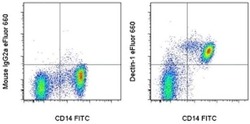
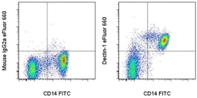
- Staining of normal human peripheral blood cells with Anti-Human CD14 FITC (Product # 11-0149-42) and Mouse IgG2a, K Isotype Control eFluor® 660 (Product # 50-4724-80) (left) or Anti-Human/Non-Human Primate CD369 (Clec7a, Dectin-1) eFluor® 660 (right). Cells in the lymphocyte and monocyte gate were used for analysis.
Supportive validation
- Submitted by
- Invitrogen Antibodies (provider)
- Main image
- Experimental details
- NULL

- Submitted by
- Invitrogen Antibodies (provider)
- Main image
- Experimental details
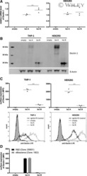
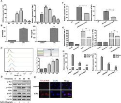
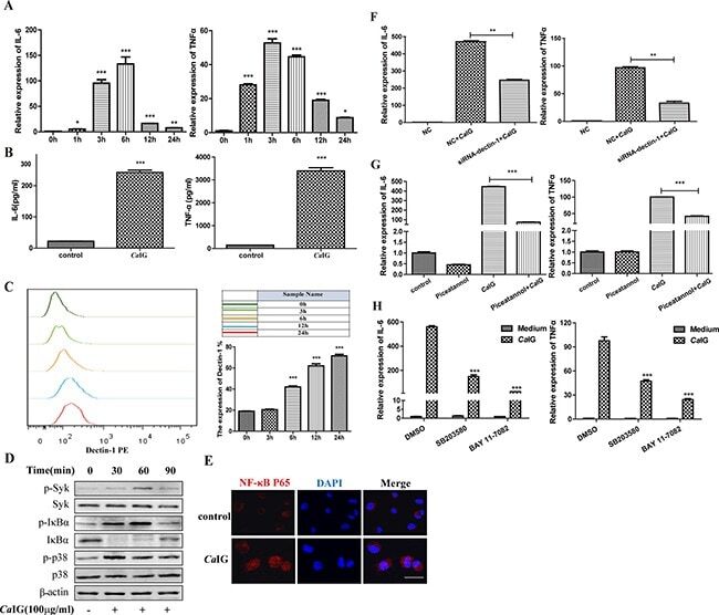
- Figure 1 Ca IG induces the transcription and expression of IL-6, TNFalpha involving the dectin-1-Syk pathways ( A ) RNA was harvested at 0, 1, 3, 6, 24 hours after treated with 100 mug/ml Ca IG and subjected to gene expression analysis by qRT-PCR normalized to beta-actin expression. For both genes analyzed, the significant difference of the Ca IG treated cells was compared with the corresponding untreated cells. ( B ) The culture medium of THP-1 cells treated with 100 mug/ml Ca IG for 24 hours was analyzed by ELISA to determine the protein level of IL-6 and TNFalpha. ( C ) The expression of dectin-1 in THP-1 cells. The THP-1 cells (2 x 10 5 cells) were incubated with anti-human primate Dectin-1 monoclonal antibody for flow cytometric analysis. ( D ) Representative images of Western Blot analyses of p-Syk, Syk, p-IkappaBalpha, IkappaBalpha, p-p38 and p38 protein levels. Protein extracts were made from untreated cells and from Ca IG treated cells for 30, 60 and 90 minutes. Extracted protein samples (50 mug per lane) were subjected to electrophoresis and immunoblotting with antibodies specific for the 6 proteins and beta-actin as control for equal loading. ( E ) NF-kappaB p65 translocation was analyzed by staining with NF-kappaB-p65 (red); and nucleuses were colored with DAPI (blue). Scale bar = 20 mum. ( F ) THP-1 cells were transfected with dectin-1 specific siRNA (siRNA-dectin-1) or scrambled control siRNA (NC) for 48 hours, and were subsequently treated with Ca IG for 6 hour
- Submitted by
- Invitrogen Antibodies (provider)
- Main image
- Experimental details
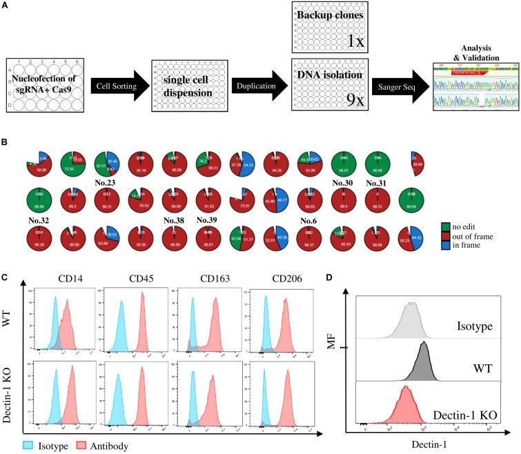
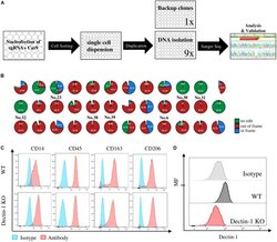
- FIGURE 5 Crispr/Cas knockout (KO) and of immune regulatory receptors in human induced pluripotent stem cells (hiPSCs) and functional analysis of macrophages derived from relative KO clones. (A) Workstream scheme for generating and analyzing of single cell KO clones in hiPSC. (B) Eight out-of-frame homozygous Dectin-1 KO clones were identified by Sanger sequencing and interference of CRISPR edits (ICE) analysis. (C) Macrophage marker expression level between wild type (WT) and Dectin-KO clone No. 32-derived macrophages. (D) Dectin-1 expression level and relative isotype in macrophages derived from WT and Dectin-1 KO clone No. 32.