Explore
 Validate
 Learn
Learn Western blot
Antibody data
- Antibody Data
- Antigen structure
- References [0]
- Comments [0]
- Validations
- Western blot [2]
- Immunohistochemistry [1]
- Flow cytometry [1]
Submit
Validation data
Reference
Comment
Report error
- Product number
- GTX81790 - Provider product page

- Provider
- GeneTex
- Proper citation
- GeneTex Cat#GTX81790, RRID:AB_11169535
- Product name
- HMGCS1 antibody, Internal
- Antibody type
- Polyclonal
- Reactivity
- Human, Mouse, Rat
- Host
- Rabbit
No comments: Submit comment
Supportive validation
- Submitted by
- GeneTex (provider)
- Main image

- Experimental details
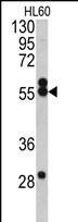
- Western blot analysis of HMGCS1 antibody (Center) (GTX81790) in HL60 cell line lysates (35μg/lane). HMGCS1 (arrow) was detected using the purified Pab.
- Submitted by
- GeneTex (provider)
- Main image

- Experimental details
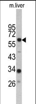
- Western blot analysis of HMGCS1 antibody (Center) (GTX81790) in mouse liver tissue lysates (35μg/lane). HMGCS1 (arrow) was detected using the purified Pab.
Supportive validation
- Submitted by
- GeneTex (provider)
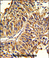
- Main image
- Experimental details
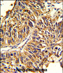
- Formalin-fixed and paraffin-embedded human lung carcinoma reacted with HMGCS1 Antibody (Center)(GTX81790), which was peroxidase-conjμgated to the secondary antibody, followed by DAB staining. This data demonstrates the use of this antibody for immunohistochemistry; clinical relevance has not been evaluated.
Supportive validation
- Submitted by
- GeneTex (provider)
- Main image

- Experimental details
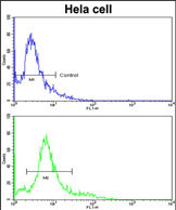
- Flow cytometric analysis of hela cells using HMGCS1 Antibody (Center)(bottom histogram)(GTX81790) compared to a negative control cell (top histogram). FITC-conjμgated goat-anti-rabbit secondary antibodies were used for the analysis.