Explore
 Validate
 Learn
Learn Western blot
 Immunocytochemistry
Antibody data
- Antibody Data
- Antigen structure
- References [0]
- Comments [0]
- Validations
- Immunocytochemistry [1]
Submit
Validation data
Reference
Comment
Report error
- Product number
- HPA049387 - Provider product page

- Provider
- Atlas Antibodies
- Proper citation
- Atlas Antibodies Cat#HPA049387, RRID:AB_2680740
- Product name
- Anti-IDH3B
- Antibody type
- Polyclonal
- Description
- Polyclonal Antibody against Human IDH3B, Gene description: isocitrate dehydrogenase 3 (NAD+) beta, Alternative Gene Names: RP46, Validated applications: ICC, IHC, WB, Uniprot ID: O43837, Storage: Store at +4°C for short term storage. Long time storage is recommended at -20°C.
- Reactivity
- Human
- Host
- Rabbit
- Conjugate
- Unconjugated
- Isotype
- IgG
- Vial size
- 100 µl
- Concentration
- 0.2 mg/ml
- Storage
- Store at +4°C for short term storage. Long time storage is recommended at -20°C.
- Handling
- The antibody solution should be gently mixed before use.
No comments: Submit comment
Supportive validation
- Submitted by
- Atlas Antibodies (provider)
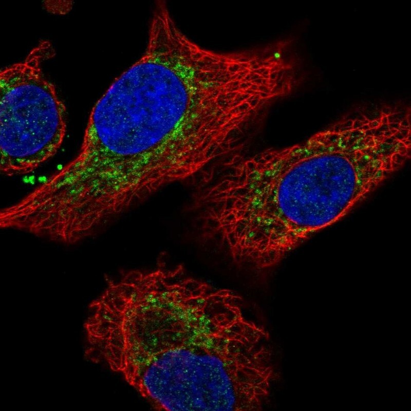
- Main image
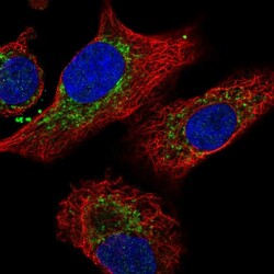
- Experimental details
- Immunofluorescent staining of human cell line A549 shows localization to mitochondria.
- Sample type
- Human