Explore
 Validate
 Learn
Learn Western blot
Antibody data
- Antibody Data
- Antigen structure
- References [0]
- Comments [0]
- Validations
- Western blot [4]
- Immunohistochemistry [2]
Submit
Validation data
Reference
Comment
Report error
- Product number
- PA5-28292 - Provider product page

- Provider
- Invitrogen Antibodies
- Product name
- HAGH Polyclonal Antibody
- Antibody type
- Polyclonal
- Antigen
- Recombinant protein fragment
- Description
- Recommended positive controls: HepG2, mouse liver. Predicted reactivity: Mouse (92%), Rat (90%), Bovine (90%). Store product as a concentrated solution. Centrifuge briefly prior to opening the vial.
- Reactivity
- Human, Mouse
- Host
- Rabbit
- Isotype
- IgG
- Vial size
- 100 µL
- Concentration
- 0.34 mg/mL
- Storage
- Store at 4°C short term. For long term storage, store at -20°C, avoiding freeze/thaw cycles.
No comments: Submit comment
Supportive validation
- Submitted by
- Invitrogen Antibodies (provider)
- Main image
- Experimental details
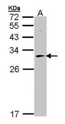
- Western blot analysis of HAGH using 50 µg of mouse liver lysate. Samples were loaded onto a 10% SDS-PAGE gel and probed with a HAGH polyclonal antibody (Product # PA5-28292) at a dilution of 1:1000.

- Submitted by
- Invitrogen Antibodies (provider)
- Main image
- Experimental details
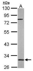
- Western blot analysis of HAGH using 30 µg of HepG2 lysate. Samples were loaded onto a 12% SDS-PAGE gel and probed with a HAGH polyclonal antibody (Product # PA5-28292) at a dilution of 1:1000.

- Submitted by
- Invitrogen Antibodies (provider)
- Main image
- Experimental details
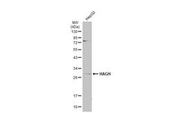
- Western Blot using HAGH Polyclonal Antibody (Product # PA5-28292). Whole cell extract (30 µg) was separated by 12% SDS-PAGE, and the membrane was blotted with HAGH Polyclonal Antibody (Product # PA5-28292) diluted at 1:1,000. The HRP-conjugated anti-rabbit IgG antibody was used to detect the primary antibody, and the signal was developed with Trident ECL plus-Enhanced.

- Submitted by
- Invitrogen Antibodies (provider)
- Main image
- Experimental details
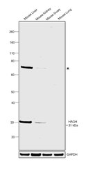
- Western blot was performed using Anti-HAGH Polyclonal Antibody (Product # PA5-28292) and a 31 kDa band corresponding to HAGH along with an uncharacterized band (*) at ~70 kDa was observed across tissue extracts tested except Mouse Ovary and Mouse Lung which are reported to be negative. Tissue extracts (30 µg lysate) of Mouse Liver (Lane 1), Mouse Kidney (Lane 2), Mouse Ovary (Lane 3) and Mouse Lung (Lane 4) were electrophoresed using NuPAGE™ 4-12% Bis-Tris Protein Gel (Product # NP0322BOX). Resolved proteins were then transferred onto a nitrocellulose membrane (Product # IB23001) by iBlot® 2 Dry Blotting System (Product # IB21001). The blot was probed with the primary antibody (1:2000 dilution) and detected by chemiluminescence with Goat anti-Rabbit IgG (H+L) Superclonal™ Recombinant Secondary Antibody, HRP (Product # A27036, 1:4000 dilution) using the iBright FL 1000 (Product # A32752). Chemiluminescent detection was performed using Novex® ECL Chemiluminescent Substrate Reagent Kit (Product # WP20005).

Supportive validation
- Submitted by
- Invitrogen Antibodies (provider)
- Main image
- Experimental details
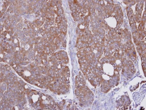
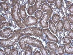
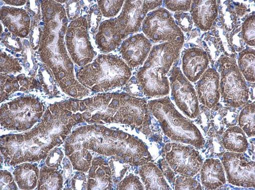
- HAGH Polyclonal Antibody detects HAGH protein at mitochondria on mouse kidney by immunohistochemical analysis. Sample: Paraffin-embedded mouse kidney. HAGH Polyclonal Antibody (Product # PA5-28292) diluted at 1:500. Antigen Retrieval: EDTA based buffer, pH 8.0, 15 min.
- Submitted by
- Invitrogen Antibodies (provider)
- Main image

- Experimental details
- Immunohistochemical analysis of paraffin-embedded NCI-N87 xenograft, using HAGH (Product # PA5-28292) antibody at 1:100 dilution. Antigen Retrieval: EDTA based buffer, pH 8.0, 15 min.