Explore
 Validate
 Learn
Learn Western blot
Antibody data
- Antibody Data
- Antigen structure
- References [1]
- Comments [0]
- Validations
- Western blot [5]
- Immunocytochemistry [2]
- Immunohistochemistry [5]
Submit
Validation data
Reference
Comment
Report error
- Product number
- PA5-78675 - Provider product page

- Provider
- Invitrogen Antibodies
- Product name
- HCN1 Polyclonal Antibody
- Antibody type
- Polyclonal
- Antigen
- Recombinant full-length protein
- Description
- Positive Control: SK-N-AS, mouse brain, rat brain Predicted Reactivity: Rabbit (90%), Bovine (89%) Store product as a concentrated solution. Centrifuge briefly prior to opening the vial.
- Reactivity
- Human, Mouse, Rat
- Host
- Rabbit
- Isotype
- IgG
- Vial size
- 100 µL
- Concentration
- 1.45 mg/mL
- Storage
- Store at 4°C short term. For long term storage, store at -20°C, avoiding freeze/thaw cycles.
Submitted references Glucocorticoid-glucocorticoid receptor-HCN1 channels reduce neuronal excitability in dorsal hippocampal CA1 neurons.
Kim J, Lei Y, Lu XY, Kim CS
Molecular psychiatry 2022 Oct;27(10):4035-4049
Molecular psychiatry 2022 Oct;27(10):4035-4049
No comments: Submit comment
Supportive validation
- Submitted by
- Invitrogen Antibodies (provider)
- Main image
- Experimental details
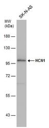
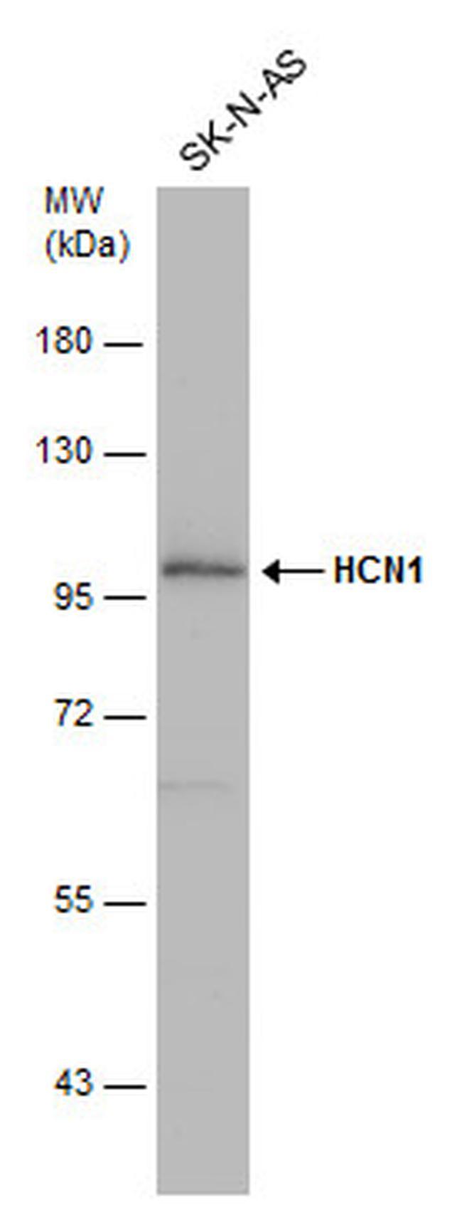
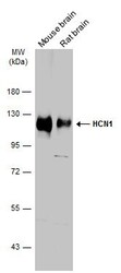
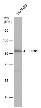
- Western blot analysis of HCN1 in whole cell lysate using 30 µg of protein. Samples were separated with 7.5% SDS-PAGE and incubated with HCN1 polyclonal antibody (Product # PA5-78675) using a dilution of 1:1000.
- Submitted by
- Invitrogen Antibodies (provider)
- Main image
- Experimental details
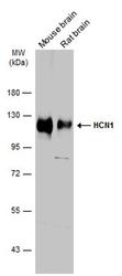
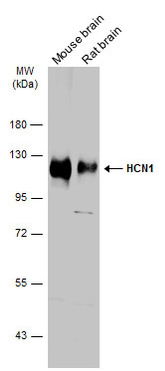
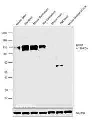
- Western blot analysis of HCN1 in various tissue extracts using 50 µg of protein. Samples were separated with 7.5% SDS-PAGE and incubated with HCN1 polyclonal antibody (Product # PA5-78675) using a dilution of 1:1000 followed by HRP-conjugated anti-rabbit IgG.
- Submitted by
- Invitrogen Antibodies (provider)
- Main image
- Experimental details
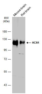
- Western Blot using HCN1 Polyclonal Antibody (Product # PA5-78675). Various tissue extracts (50 µg) were separated by 7.5% SDS-PAGE, and the membrane was blotted with HCN1 Polyclonal Antibody (Product # PA5-78675) diluted at 1:1,000. The HRP-conjugated anti-rabbit IgG antibody was used to detect the primary antibody.
- Submitted by
- Invitrogen Antibodies (provider)
- Main image
- Experimental details
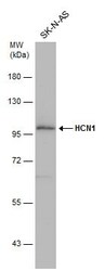
- Western Blot using HCN1 Polyclonal Antibody (Product # PA5-78675). Whole cell extract (30 µg) was separated by 7.5% SDS-PAGE, and the membrane was blotted with HCN1 Polyclonal Antibody (Product # PA5-78675) diluted at 1:1,000.
- Submitted by
- Invitrogen Antibodies (provider)
- Main image
- Experimental details
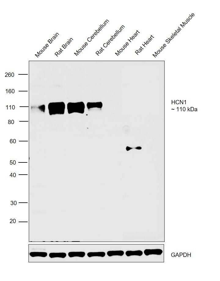
- Western blot was performed using Anti-HCN1 Polyclonal Antibody (Product # PA5-78675) and a 110 kDa band corresponding to Potassium/sodium hyperpolarization-activated cyclic nucleotide-gated channel 1 was observed only in Mouse Brain, Rat Brain, Mouse Cerebellum and Rat cerebellum but not in any other tissues tested like Mouse Heart, Rat Heart and Mouse Skeletal Muscle. Tissue extracts (30 µg lysate) of Mouse Brain (Lane 1), Rat Brain (Lane 2), Mouse Cerebellum (Lane 3), Rat Cerebellum (Lane 4), Mouse Heart (Lane 5), Rat Heart (Lane 6) and Mouse Skeletal Muscle (Lane 7) were electrophoresed using NuPAGE™ 4-12% Bis-Tris Protein Gel (Product # NP0322BOX). Resolved proteins were then transferred onto a nitrocellulose membrane (Product # IB23001) by iBlot® 2 Dry Blotting System (Product # IB21001). The blot was probed with the primary antibody (1:1000 dilution) and detected by chemiluminescence with Goat anti-Rabbit IgG (H+L) Superclonal™ Recombinant Secondary Antibody, HRP (Product # A27036,1:4000 dilution) using the iBright FL 1000 (Product # A32752). Chemiluminescent detection was performed using Novex® ECL Chemiluminescent Substrate Reagent Kit (Product # WP20005).
Supportive validation
- Submitted by
- Invitrogen Antibodies (provider)
- Main image
- Experimental details
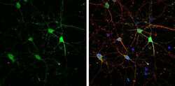
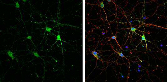
- Immunocytochemistry-Immunofluorescence analysis of HCN1 was performed in DIV9 rat E18 primary cortical neuron cells fixed in 4% paraformaldehyde at RT for 15 min. Green: HCN1 Polyclonal Antibody (Product # PA5-78675) diluted at 1:500. Red: beta Tubulin 3/ Tuj1, stained by beta Tubulin 3/ Tuj1 antibody. Blue: Fluoroshield with DAPI.
- Submitted by
- Invitrogen Antibodies (provider)
- Main image
- Experimental details
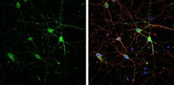
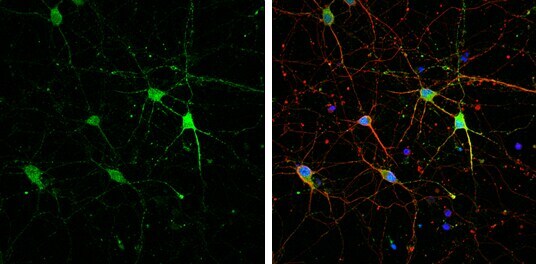
- Immunocytochemistry-Immunofluorescence analysis of HCN1 was performed in DIV9 rat E18 primary cortical neuron cells fixed in 4% paraformaldehyde at RT for 15 min. Green: HCN1 Polyclonal Antibody (Product # PA5-78675) diluted at 1:500. Red: beta Tubulin 3/ Tuj1, stained by beta Tubulin 3/ Tuj1 antibody. Blue: Fluoroshield with DAPI.
Supportive validation
- Submitted by
- Invitrogen Antibodies (provider)
- Main image
- Experimental details
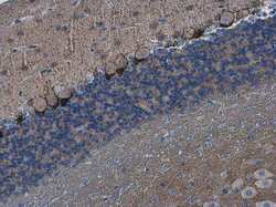
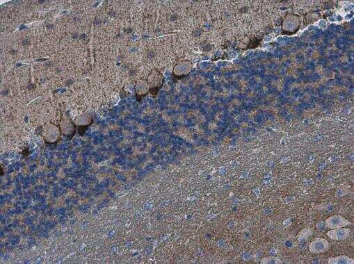
- HCN1 Polyclonal Antibody detects HCN1 protein at cell membrane in mouse brain by immunohistochemical analysis. Sample: Paraffin-embedded mouse brain. HCN1 Polyclonal Antibody (Product # PA5-78675) diluted at 1:500. Antigen Retrieval: Citrate buffer, pH 6.0, 15 min.
- Submitted by
- Invitrogen Antibodies (provider)
- Main image
- Experimental details
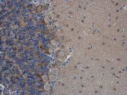
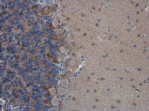
- HCN1 Polyclonal Antibody detects HCN1 protein at cell membrane in rat brain by immunohistochemical analysis. Sample: Paraffin-embedded rat brain. HCN1 Polyclonal Antibody (Product # PA5-78675) diluted at 1:500. Antigen Retrieval: Citrate buffer, pH 6.0, 15 min.
- Submitted by
- Invitrogen Antibodies (provider)
- Main image
- Experimental details
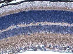
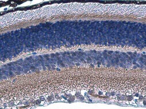
- HCN1 Polyclonal Antibody detects HCN1 protein in mouse retina by immunohistochemical analysis. Sample: Paraffin-embedded mouse retina. HCN1 Polyclonal Antibody (Product # PA5-78675) diluted at 1:800. Antigen Retrieval: Citrate buffer, pH 6.0, 15 min.
- Submitted by
- Invitrogen Antibodies (provider)
- Main image
- Experimental details
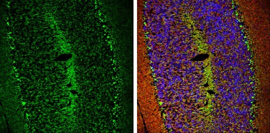
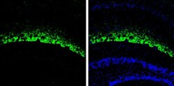
- HCN1 Polyclonal Antibody detects HCN1 protein expression by immunohistochemical analysis. Sample: Frozen-sectioned adult mouse hippocampus. Green: HCN1 protein stained by HCN1 Polyclonal Antibody (Product # PA5-78675) diluted at 1:250. Blue: Fluoroshield with DAPI .

- Submitted by
- Invitrogen Antibodies (provider)
- Main image

- Experimental details
- HCN1 Polyclonal Antibody detects HCN1 protein expression by immunohistochemical analysis. Sample: Frozen-sectioned adult mouse cerebellum. Green: HCN1 protein stained by HCN1 Polyclonal Antibody (Product # PA5-78675) diluted at 1:250. Red: beta Tubulin 3/ TUJ1, stained by beta Tubulin 3/ TUJ1 antibody [GT11710] diluted at 1:500. Blue: Fluoroshield with DAPI .