 Explore
 Validate
 Learn
Learn Western blot
 ELISA
ELISAAntibody data
- Antibody Data
- Antigen structure
- References [9]
- Comments [0]
- Validations
- Western blot [1]
- Immunocytochemistry [1]
- Immunohistochemistry [2]
Submit
Validation data
Reference
Comment
Report error
- Product number
- 14524-1-AP - Provider product page

- Provider
- Proteintech Group
- Proper citation
- Proteintech Cat#14524-1-AP, RRID:AB_2148137
- Product name
- MYBBP1A antibody
- Antibody type
- Polyclonal
- Description
- KD/KO validated MYBBP1A antibody (Cat. #14524-1-AP) is a rabbit polyclonal antibody that shows reactivity with human and has been validated for the following applications: FC, IF, IHC, IP, WB,ELISA.
- Reactivity
- Human
- Host
- Rabbit
- Conjugate
- Unconjugated
- Isotype
- IgG
- Vial size
- 20ul, 150ul
Submitted references Analysis of the Porcine Reproductive and Respiratory Syndrome Virus Nucleocapsid Interactome.
USP30-AS1 Suppresses Colon Cancer Cell Inflammatory Response Through NF-κB/MYBBP1A Signaling.
Nuclear reorganization by NPM1-mediated phase separation triggered by adenovirus core protein VII.
Non-immune hydrops fetalis is associated with bi-allelic pathogenic variants in the MYB Binding Protein 1a (MYBBP1A) gene.
The nucleolar phase of signal recognition particle assembly.
A fetal tumor suppressor axis abrogates MLL-fusion-driven acute myeloid leukemia.
In vivo protein turnover rates in varying oxygen tensions nominate MYBBP1A as a mediator of the hyperoxia response.
c-MYB- and PGC1a-dependent metabolic switch induced by MYBBP1A loss in renal cancer.
Loss of MYBBP1A Induces Cancer Stem Cell Activity in Renal Cancer.
Kovanich D, Ketsuwan K, Hengphasatporn K, Thepparit C, Sittipaisankul P, Wongkongkathep P, Sirisereewan C, Techakriengkrai N, Nedumpun T, Shigeta Y, Pisitkun T, Suradhat S
Journal of proteome research 2025 Nov 7;24(11):5390-5411
Journal of proteome research 2025 Nov 7;24(11):5390-5411
USP30-AS1 Suppresses Colon Cancer Cell Inflammatory Response Through NF-κB/MYBBP1A Signaling.
Wang R, Li X, Jiang Y, Zhang H, Yang S, Xie W, Xu N
Inflammation 2025 Aug;48(4):1998-2008
Inflammation 2025 Aug;48(4):1998-2008
Nuclear reorganization by NPM1-mediated phase separation triggered by adenovirus core protein VII.
Genoveso MJ, Okuwaki M, Kato K, Nagata K, Kawaguchi A
Microbiology spectrum 2024 Oct 3;12(10):e0041624
Microbiology spectrum 2024 Oct 3;12(10):e0041624
Non-immune hydrops fetalis is associated with bi-allelic pathogenic variants in the MYB Binding Protein 1a (MYBBP1A) gene.
Tenorio-Castano J, Mansilla Aparicio E, García Santiago FA, Klotz CM, Regojo RM, Anguita E, Ryan E, Juusola J, Herrero B, Arias P, Parra A, Pascual P, Gallego N, Cazalla M, Rodriguez-González R, Antolín E, Nevado J, Ruiz-Perez VL, Lapunzina P
Clinical genetics 2024 Dec;106(6):713-720
Clinical genetics 2024 Dec;106(6):713-720
The nucleolar phase of signal recognition particle assembly.
Issa A, Schlotter F, Flayac J, Chen J, Wacheul L, Philippe M, Sardini L, Mostefa L, Vandermoere F, Bertrand E, Verheggen C, Lafontaine DL, Massenet S
Life science alliance 2024 Aug;7(8)
Life science alliance 2024 Aug;7(8)
A fetal tumor suppressor axis abrogates MLL-fusion-driven acute myeloid leukemia.
Eldeeb M, Yuan O, Guzzi N, Thi Ngoc PC, Konturek-Ciesla A, Kristiansen TA, Muthukumar S, Magee J, Bellodi C, Yuan J, Bryder D
Cell reports 2023 Feb 28;42(2):112099
Cell reports 2023 Feb 28;42(2):112099
In vivo protein turnover rates in varying oxygen tensions nominate MYBBP1A as a mediator of the hyperoxia response.
Chen X, Haribowo AG, Baik AH, Fossati A, Stevenson E, Chen YR, Reyes NS, Peng T, Matthay MA, Traglia M, Pico AR, Jarosz DF, Buchwalter A, Ghaemmaghami S, Swaney DL, Jain IH
Science advances 2023 Dec 8;9(49):eadj4884
Science advances 2023 Dec 8;9(49):eadj4884
c-MYB- and PGC1a-dependent metabolic switch induced by MYBBP1A loss in renal cancer.
Felipe-Abrio B, Verdugo-Sivianes EM, Carnero A
Molecular oncology 2019 Jul;13(7):1519-1533
Molecular oncology 2019 Jul;13(7):1519-1533
Loss of MYBBP1A Induces Cancer Stem Cell Activity in Renal Cancer.
Felipe-Abrio B, Verdugo-Sivianes EM, Sáez C, Carnero A
Cancers 2019 Feb 18;11(2)
Cancers 2019 Feb 18;11(2)
No comments: Submit comment
Supportive validation
- Submitted by
- Proteintech Group (provider)
- Main image
- Experimental details
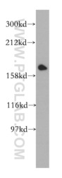
- HeLa cells were subjected to SDS PAGE followed by western blot with 14524-1-AP(MYBBP1A antibody) at dilution of 1:500
- Sample type
- cell line

Supportive validation
- Submitted by
- Proteintech Group (provider)
- Main image
- Experimental details
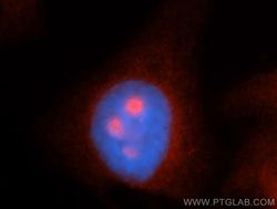
- Immunofluorescent analysis of HepG2 cells, using MYBBP1A antibody 14524-1-AP at 1:50 dilution and Rhodamine-labeled goat anti-rabbit IgG (red). Blue pseudocolor = DAPI (fluorescent DNA dye).
- Sample type
- cell line
Supportive validation
- Submitted by
- Proteintech Group (provider)
- Main image

- Experimental details
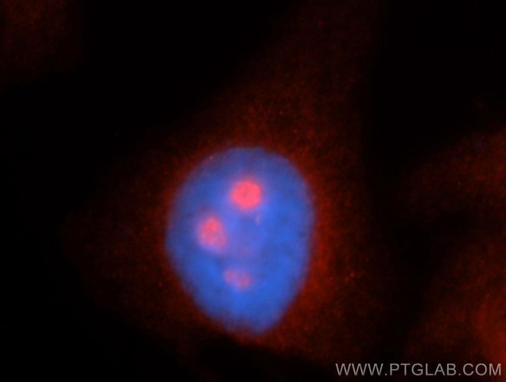
- Immunohistochemical of paraffin-embedded human kidney using 14524-1-AP(MYBBP1A antibody) at dilution of 1:100 (under 10x lens)
- Sample type
- tissue
- Submitted by
- Proteintech Group (provider)
- Main image
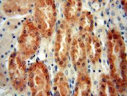
- Experimental details
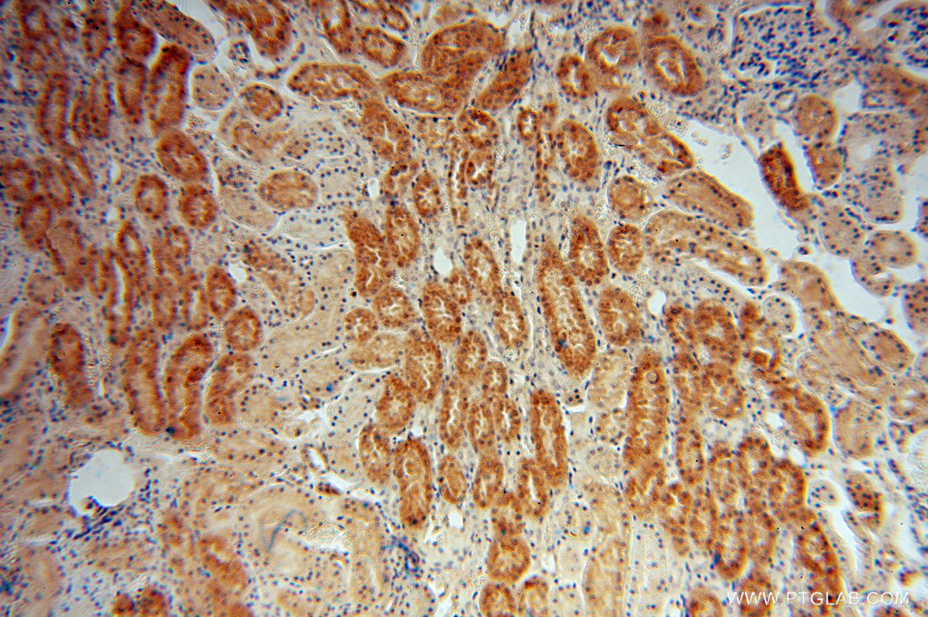
- Immunohistochemical of paraffin-embedded human kidney using 14524-1-AP(MYBBP1A antibody) at dilution of 1:100 (under 40x lens)
- Sample type
- tissue
