Explore
 Validate
 Learn
Learn Western blot
Antibody data
- Antibody Data
- Antigen structure
- References [1]
- Comments [0]
- Validations
- Western blot [2]
- Immunocytochemistry [2]
- Immunohistochemistry [3]
- Flow cytometry [1]
- Other assay [1]
Submit
Validation data
Reference
Comment
Report error
- Product number
- PA5-18221 - Provider product page

- Provider
- Invitrogen Antibodies
- Product name
- ORP5 Polyclonal Antibody
- Antibody type
- Polyclonal
- Antigen
- Synthetic peptide
- Description
- This antibody is predicted to react with canine, mouse and rat based on sequence homology. This antibody is tested in Peptide ELISA: antibody detection limit dilution 32,000.
- Reactivity
- Human, Rat
- Host
- Goat
- Isotype
- IgG
- Vial size
- 100 μg
- Concentration
- 0.5 mg/mL
- Storage
- -20°C, Avoid Freeze/Thaw Cycles
Submitted references 2-Hydroxypropyl-gamma-cyclodextrin overcomes NPC1 deficiency by enhancing lysosome-ER association and autophagy.
Singhal A, Krystofiak ES, Jerome WG, Song B
Scientific reports 2020 May 26;10(1):8663
Scientific reports 2020 May 26;10(1):8663
No comments: Submit comment
Supportive validation
- Submitted by
- Invitrogen Antibodies (provider)
- Main image
- Experimental details
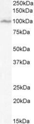
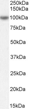
- Western blot analysis of ORP5 using ORP5 Polyclonal Antibody (Product # PA5-18221) (0.1 µg/mL) in staining of Jurkat cell lysate (35 µg protein in RIPA buffer). Detected by chemiluminescence.

- Submitted by
- Invitrogen Antibodies (provider)
- Main image
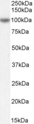
- Experimental details
- Western blot analysis of ORP5 using ORP5 Polyclonal Antibody (Product # PA5-18221) (0.3 µg/mL) in staining of Rat Spleen lysate (35 µg protein in RIPA buffer). Detected by chemiluminescence.
Supportive validation
- Submitted by
- Invitrogen Antibodies (provider)
- Main image

- Experimental details
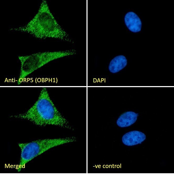
- Immunocytochemistry analysis of ORP5 using ORP5 Polyclonal Antibody (Product # PA5-18221) in paraformaldehyde fixed HeLa cells, permeabilized with 0.15% Triton. Primary incubation 1hr (10 µg/mL) followed by Alexa Fluor 488 secondary antibody (2 µg/mL), showing cytoplasmic staining. The nuclear stain is DAPI (blue). Negative control: Unimmunized goat IgG (10 µg/mL) followed by Alexa Fluor 488 secondary antibody (2 µg/mL).
- Submitted by
- Invitrogen Antibodies (provider)
- Main image
- Experimental details
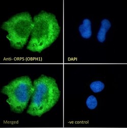
- Immunocytochemistry analysis of ORP5 using ORP5 Polyclonal Antibody (Product # PA5-18221) in paraformaldehyde fixed U2OS cells, permeabilized with 0.15% Triton. Primary incubation 1hr (10 µg/mL) followed by Alexa Fluor 488 secondary antibody (2 µg/mL), showing cytoplasmic staining. The nuclear stain is DAPI (blue). Negative control: Unimmunized goat IgG (10 µg/mL) followed by Alexa Fluor 488 secondary antibody (2 µg/mL).

Supportive validation
- Submitted by
- Invitrogen Antibodies (provider)
- Main image
- Experimental details
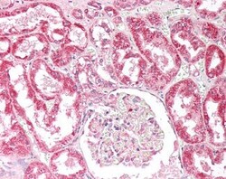
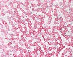
- Immunohistochemistry analysis of ORP5 in Human Kidney (Formalin-Fixed, Paraffin-Embedded). Samples were incubated with ORP5 polyclonal antibody (Product # PA5-18221).
- Submitted by
- Invitrogen Antibodies (provider)
- Main image
- Experimental details
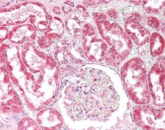
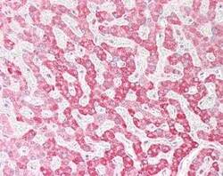
- Immunohistochemistry analysis of ORP5 in Human Liver (Formalin-Fixed, Paraffin-Embedded). Samples were incubated with ORP5 polyclonal antibody (Product # PA5-18221).

- Submitted by
- Invitrogen Antibodies (provider)
- Main image
- Experimental details
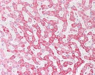
- Immunohistochemistry analysis of ORP5 in Human Liver (Formalin-Fixed, Paraffin-Embedded). Samples were incubated with ORP5 polyclonal antibody (Product # PA5-18221).
Supportive validation
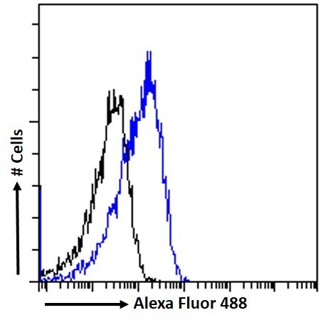
- Submitted by
- Invitrogen Antibodies (provider)
- Main image
- Experimental details
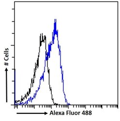
- Flow Cytometry analysis of ORP5 using ORP5 Polyclonal Antibody (Product # PA5-18221) in paraformaldehyde fixed HeLa cells (blue line), permeabilized with 0.5% Triton. Primary incubation 1hr (10 µg/mL) followed by Alexa Fluor 488 secondary antibody (1 µg/mL). IgG control: Unimmunized goat IgG (black line) followed by Alexa Fluor 488 secondary antibody.
Supportive validation
- Submitted by
- Invitrogen Antibodies (provider)
- Main image
- Experimental details
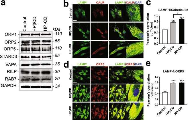
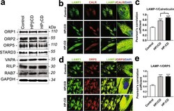
- Figure 5 HPgammaCD enhances lysosome-ER association in NPC1 fibroblasts. NPC1 mutant cells were treated with HPgammaCD or HPbetaCD (1 mM, 72 h). Cells were then analyzed for the levels of lysosome and ER contact proteins by immunoblotting ( a ) and for lysosome-ER association by confocal microscopy ( b-e ). The blots are from different parts of the same gel and delineated with dividing lines. Confocal microscopy was used to visualize the lysosome and ER by immunostaining cells for the lysosomal membrane protein (LAMP1, green) and the ER protein (Calreticulin or ORP5, red). Microscopic images showed co-localization of LAMP1 and CALR ( b ) or ORP5 ( d ) as a yellow color (merge). Nuclei were stained using DAPI (blue). The co-localization, measured by Pearson's correlation coefficient, was significantly higher in the CD-treated cells compared to untreated control cells ( c,e ). Data are mean +- S.E.M. of triplicates and a representative of three independent experiments. Symbols indicate the relative level of significance compared with the control (*P < 0.05, ***P < 0.001). Scale bar = 50 um.