Explore
 Validate
 Learn
Learn Western blot
 ELISA
ELISA Immunocytochemistry
Antibody data
- Antibody Data
- Antigen structure
- References [3]
- Comments [0]
- Validations
- Immunocytochemistry [2]
- Immunohistochemistry [1]
- Other assay [2]
Submit
Validation data
Reference
Comment
Report error
- Product number
- MA5-15515 - Provider product page

- Provider
- Invitrogen Antibodies
- Product name
- ISL1 Monoclonal Antibody (1H9)
- Antibody type
- Monoclonal
- Antigen
- Purifed from natural sources
- Description
- MA5-15515 targets ISL1 in indirect ELISA, IF, IHC, and WB applications and shows reactivity with Human samples. The MA5-15515 immunogen is purified recombinant fragment of human ISL1 expressed in E. Coli. MA5-15515 detects ISL1 which has a predicted molecular weight of approximately 39kDa.
- Reactivity
- Human
- Host
- Mouse
- Isotype
- IgG
- Antibody clone number
- 1H9
- Vial size
- 100 μL
- Concentration
- Conc. not determined
- Storage
- Store at 4°C short term. For long term storage, store at -20°C, avoiding freeze/thaw cycles.
Submitted references Comparison of induced neurons reveals slower structural and functional maturation in humans than in apes.
Herpes simplex virus type 1 infection leads to neurodevelopmental disorder-associated neuropathological changes.
Engineering stem cell-derived 3D brain organoids in a perfusable organ-on-a-chip system.
Schörnig M, Ju X, Fast L, Ebert S, Weigert A, Kanton S, Schaffer T, Nadif Kasri N, Treutlein B, Peter BM, Hevers W, Taverna E
eLife 2021 Jan 20;10
eLife 2021 Jan 20;10
Herpes simplex virus type 1 infection leads to neurodevelopmental disorder-associated neuropathological changes.
Qiao H, Guo M, Shang J, Zhao W, Wang Z, Liu N, Li B, Zhou Y, Wu Y, Chen P
PLoS pathogens 2020 Oct;16(10):e1008899
PLoS pathogens 2020 Oct;16(10):e1008899
Engineering stem cell-derived 3D brain organoids in a perfusable organ-on-a-chip system.
Wang Y, Wang L, Guo Y, Zhu Y, Qin J
RSC advances 2018 Jan 2;8(3):1677-1685
RSC advances 2018 Jan 2;8(3):1677-1685
No comments: Submit comment
Supportive validation
- Submitted by
- Invitrogen Antibodies (provider)
- Main image
- Experimental details
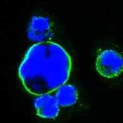
- Immunofluorescence analysis of HEK293 cells trasfected with full-length ISL1-human IgG Fc using ISL1 monoclonal antibody (Product # MA5-15515) (Green). Blue: DRAQ5 fluorescent DNA dye.

- Submitted by
- Invitrogen Antibodies (provider)
- Main image
- Experimental details
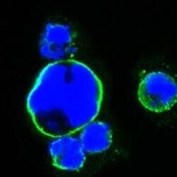
- Immunofluorescence analysis of HEK293 cells trasfected with full-length ISL1-human IgG Fc using ISL1 monoclonal antibody (Product # MA5-15515) (Green). Blue: DRAQ5 fluorescent DNA dye.

Supportive validation
- Submitted by
- Invitrogen Antibodies (provider)
- Main image

- Experimental details
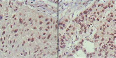
- Immunohistochemical analysis of paraffin-embedded human lung cancer (left) and cervical carcinoma (right) using ISL1 monoclonal antibody (Product # MA5-15515) followed with DAB staining.
Supportive validation
- Submitted by
- Invitrogen Antibodies (provider)
- Main image

- Experimental details
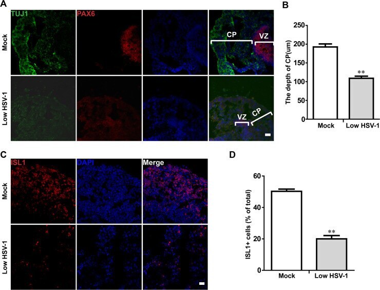
- Fig 5 Dysregulated the cortical layer and hindbrain populations in HSV-1-infected neuroepithelial buds. (A) The dorsal NSC marker (PAX6) and newborn neuron marker (TUJ) were observed in the brain cerebral organoids at the 18 day after 3 days with HSV-1 infection (Mock, Low infection). Scale bars: 25 mum. The images were taken Images were captured on a Leica TCS SP8 STED confocal microscope. (B) Comparison of depths of CP layer between mock- and HSV-1-infected neuroepithelial buds. (C) Immunofluorescence staining for the hindbrain marker (ISL1) in neuroepithelial buds at the day 18 after 3 days with HSV-1 infection. Scale bars: 25 mum. (D) Quantification of ISL1-positive cells in the presence or absence of HSV-1 infection. Data represent the mean +- SEM from four experiments. **p < 0.01 by Student's t test.
- Submitted by
- Invitrogen Antibodies (provider)
- Main image
- Experimental details
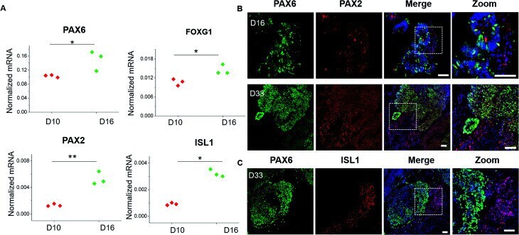
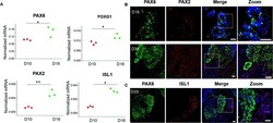
- Identification of specific brain regions in brain organoids. (A) Forebrain markers (PAX6, FOXG1) and hindbrain markers (PAX2, ISL1) were quantified by RT-PCR in spheroids at 10 and 16 days of differentiation. The expression values were normalized to the beta-actin expression level. Three independent experiments had been performed. Data are shown as mean +- SD. * p < 0.05, ** p < 0.01. (B) Immunohistochemical staining of PAX6 and PAX2 markers in cryosections of spheroids at 16 days and 33 days. (C) Immunohistochemical staining of PAX6 and ISL1 markers in cryosections of brain organoids at 33 days. Scale bars = 50 mum.