Explore
 Validate
 Learn
Learn Western blot
Antibody data
- Antibody Data
- Antigen structure
- References [0]
- Comments [0]
- Validations
- Western blot [6]
- Immunocytochemistry [2]
- Immunohistochemistry [2]
Submit
Validation data
Reference
Comment
Report error
- Product number
- PA5-27789 - Provider product page

- Provider
- Invitrogen Antibodies
- Product name
- ISL1 Polyclonal Antibody
- Antibody type
- Polyclonal
- Antigen
- Recombinant protein fragment
- Description
- Recommended positive controls: K562 whole cell extract, K562 nuclear extract, jurkat whole cell extract, jurkat nuclear extract, PC-12. Predicted reactivity: Mouse (100%), Rat (100%), Zebrafish (97%), Japanese Medaka (97%), Xenopus laevis (94%), Chicken (99%), Bovine (100%). Store product as a concentrated solution. Centrifuge briefly prior to opening the vial.
- Reactivity
- Human, Mouse, Rat
- Host
- Rabbit
- Isotype
- IgG
- Vial size
- 100 µL
- Concentration
- 0.55 mg/mL
- Storage
- Store at 4°C short term. For long term storage, store at -20°C, avoiding freeze/thaw cycles.
No comments: Submit comment
Supportive validation
- Submitted by
- Invitrogen Antibodies (provider)
- Main image
- Experimental details
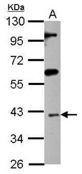
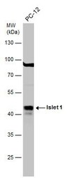
- Western blot analysis of Islet 1 using 30 µg of HL-60 lysate. Samples were loaded onto a 10% SDS-PAGE gel and probed with an Islet 1 polyclonal antibody (Product # PA5-27789) at a dilution of 1:1000.

- Submitted by
- Invitrogen Antibodies (provider)
- Main image
- Experimental details
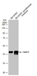
- Western Blot analysis of ISL1 was performed by separating 30 µg of SH-SY5Y whole cell and nuclear extracts by 10% SDS-PAGE. Proteins were transferred to a membrane and probed with a ISL1 Polyclonal Antibody (Product # PA5-27789) at a dilution of 1:10000. The HRP-conjugated anti-rabbit IgG antibody was used to detect the primary antibody.

- Submitted by
- Invitrogen Antibodies (provider)
- Main image
- Experimental details
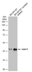
- Western Blot using ISL1 Polyclonal Antibody (Product # PA5-27789). SH-SY5Y whole cell and nuclear extracts (30 µg) were separated by 10% SDS-PAGE, and the membrane was blotted with ISL1 Polyclonal Antibody (Product # PA5-27789) diluted at 1:10,000. The HRP-conjugated anti-rabbit IgG antibody was used to detect the primary antibody.

- Submitted by
- Invitrogen Antibodies (provider)
- Main image
- Experimental details
- Western blot analysis of ISL1 was performed by separating 30 µg of whole cell extract by 10% SDS-PAGE. Proteins were transferred to a membrane and probed with a ISL1 Polyclonal Antibody (Product # PA5-27789) at a dilution of 1:1000. The HRP-conjugated anti-rabbit IgG antibody was used to detect the primary antibody.

- Submitted by
- Invitrogen Antibodies (provider)
- Main image
- Experimental details
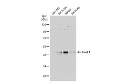
- Western Blot using ISL1 Polyclonal Antibody (Product # PA5-27789). Various whole cell extracts (30 µg) were separated by 10% SDS-PAGE, and the membrane was blotted with ISL1 Polyclonal Antibody (Product # PA5-27789) diluted at 1:10,000. The HRP-conjugated anti-rabbit IgG antibody was used to detect the primary antibody.

- Submitted by
- Invitrogen Antibodies (provider)
- Main image
- Experimental details
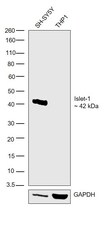
- Western blot was performed using Anti-ISL1 Rabbit Polyclonal Antibody (Product # PA5-27789) and a 42 kDa band corresponding to Islet-1 was observed in SH-SY5Y but not in THP1 cells. Whole cell extracts (30 µg lysate) of SH-SY5Y (Lane 1) or THP1 (Lane 2) were electrophoresed using NuPAGE® 4-12 % Bis-Tris gel (Product # NP0322BOX). Resolved proteins were then transferred onto a nitrocellulose membrane (Product # IB23001) by iBlot® 2 Dry Blotting System (Product # IB21001).The blot was probed with the primary antibody (1:1000 dilution) and detected by chemiluminescence with Goat anti-Rabbit IgG (H+L) Superclonal™ Recombinant Secondary Antibody, HRP (Product # A27036, 1:4000 dilution) using the iBright FL 1000 (Product # A32752). Chemiluminescent detection was performed using Novex® ECL Chemiluminescent Substrate Reagent Kit (Product # WP20005).

Supportive validation
- Submitted by
- Invitrogen Antibodies (provider)
- Main image

- Experimental details
- ISL1 Polyclonal Antibody detects Islet 1 protein at nucleus by immunofluorescent analysis. Sample: SK-N-AS cells were fixed in 4% paraformaldehyde at RT for 15 min. Green: Islet 1 protein stained by ISL1 Polyclonal Antibody (Product # PA5-27789) diluted at 1:500. Blue: Hoechst 33342 staining. Scale bar = 10 µm.

- Submitted by
- Invitrogen Antibodies (provider)
- Main image

- Experimental details
- Immunofluorescence analysis of ISL1 was performed using 70% confluent log phase SH-SY5Y cells. The cells were fixed with 4% paraformaldehyde for 10 minutes, permeabilized with 0.1% Triton™ X-100 for 15 minutes, and blocked with 2% BSA for 1 hour at room temperature. The cells were labeled with ISL1 Polyclonal Antibody (Product # PA5-27789) at 1:100 dilution in 0.1% BSA, incubated at 4 degree Celsius overnight and then labeled with Goat anti-Rabbit IgG (H+L) Superclonal™ Recombinant Secondary Antibody, Alexa Fluor® 488 (Product # A27034) at a dilution of 1:2000 for 45 minutes at room temperature (Panel a: green). Nuclei (Panel b: blue) were stained with ProLong™ Diamond Antifade Mountant with DAPI (Product # P36962). F-actin (Panel c: red) was stained with Rhodamine Phalloidin (Product # R415). Panel d represents the merged image showing nuclear localization. Panel e represents control cells with no primary antibody to assess background. The images were captured at 60X magnification.

Supportive validation
- Submitted by
- Invitrogen Antibodies (provider)
- Main image
- Experimental details
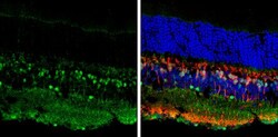
- Immunohistochemistry (Frozen) analysis of ISL1 was performed in frozen sectioned adult mouse retina tissue using ISL1 Polyclonal Antibody (Product # PA5-27789) at a dilution of 1:250 (Green). Red: Protein kinase C alpha staining. Blue: Fluoroshield with DAPI.

- Submitted by
- Invitrogen Antibodies (provider)
- Main image
- Experimental details
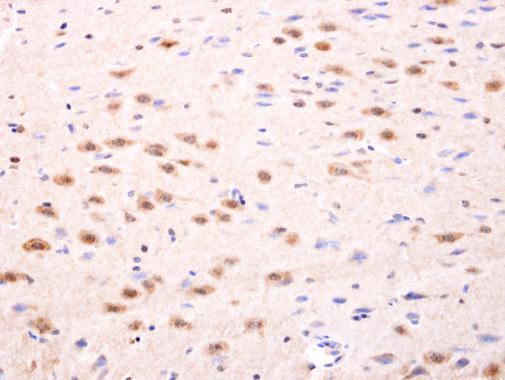
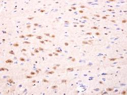
- ISL1 Polyclonal Antibody detects Islet 1 protein at nucleus on rat fore brain by immunohistochemical analysis. Sample: Paraffin-embedded rat fore brain. ISL1 Polyclonal Antibody (Product # PA5-27789) dilution: 1:100. Antigen Retrieval: EDTA based buffer, pH 8.0, 15 min.