 Explore
 Validate
 Learn
Learn Western blot
Antibody data
- Antibody Data
- Antigen structure
- References [0]
- Comments [0]
- Validations
- Western blot [3]
- Immunocytochemistry [4]
- Immunohistochemistry [1]
Submit
Validation data
Reference
Comment
Report error
- Product number
- PA5-59051 - Provider product page

- Provider
- Invitrogen Antibodies
- Product name
- NDUFAF1 Polyclonal Antibody
- Antibody type
- Polyclonal
- Antigen
- Recombinant protein fragment
- Description
- Immunogen sequence: LLKDEIVDHW RGPEGHPLHE VLLEQAKVVW QFRGKEDLDK WTVTSDKTIG GRSEVFLKMG KNNQSALLYG TLSSEAPQDG ESTRSGYCAM ISRIPRGAF Highest antigen sequence identity to the following orthologs: Mouse - 82%, Rat - 84%.
- Reactivity
- Human
- Host
- Rabbit
- Isotype
- IgG
- Vial size
- 100 μL
- Concentration
- 0.6 mg/mL
- Storage
- Store at 4°C short term. For long term storage, store at -20°C, avoiding freeze/thaw cycles.
No comments: Submit comment
Supportive validation
- Submitted by
- Invitrogen Antibodies (provider)
- Main image

- Experimental details
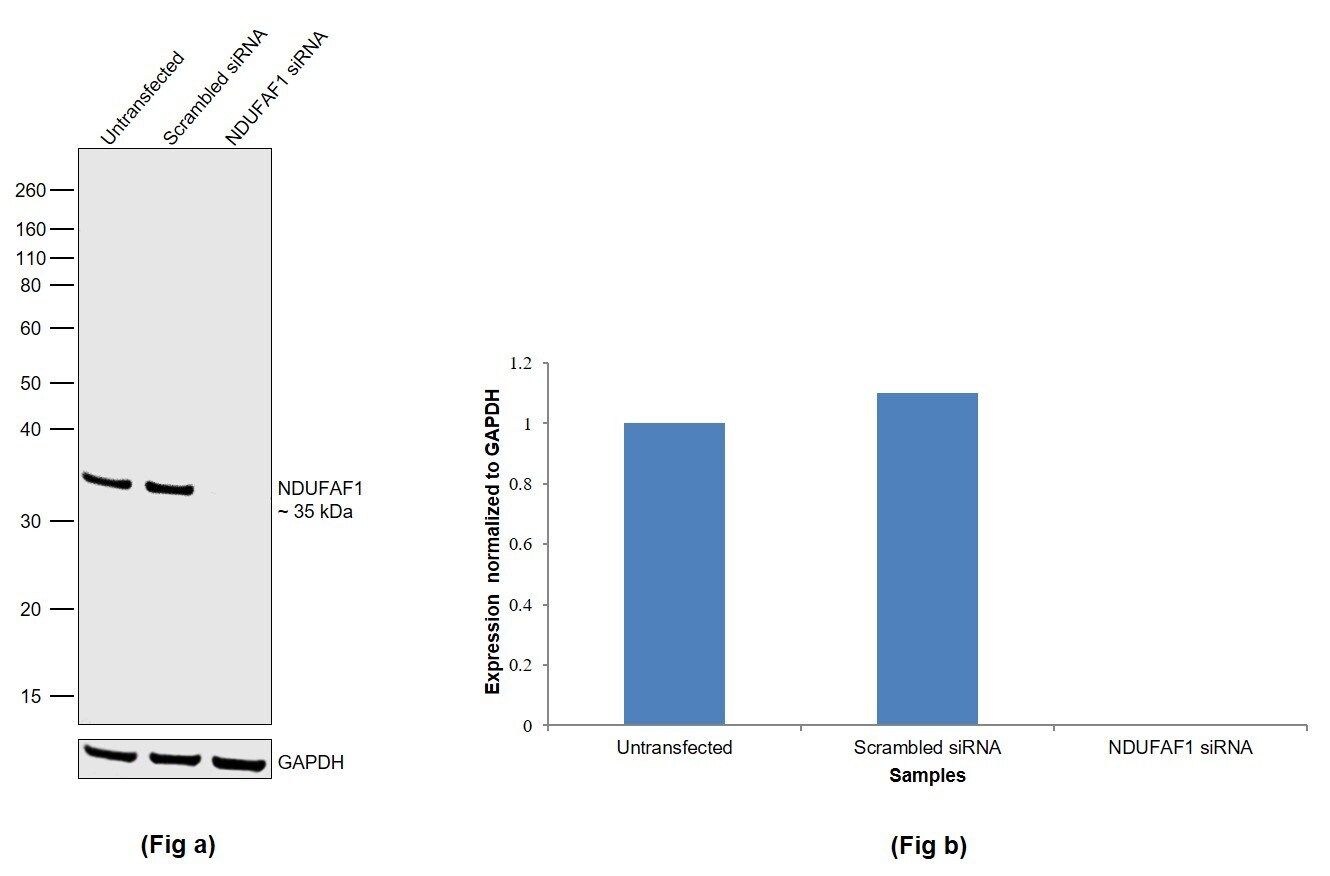
- Knockdown of Complex I intermediate-associated protein 30, mitochondrial was achieved by transfecting HEK-293 with Complex I intermediate-associated protein 30, mitochondrial specific siRNAs (Silencer® select Product # S27439, S27438). Western Blot analysis (Fig. a) was performed using Whole cell extracts from the Complex I intermediate-associated protein 30, mitochondrial knockdown cells (lane 3), non-targeting scrambled siRNA transfected cells (lane 2) and untransfected cells (lane 1). The blot was probed with NDUFAF1 Polyclonal Antibody (Product # PA5-59051, 0.2 µg/mL ) and Goat anti-Rabbit IgG (Heavy Chain) Superclonal™ Recombinant Secondary Antibody, HRP (Product # A27036, 1:10000). Densitometric analysis of this Western Blot is shown in histogram (Fig. b). Decrease in signal upon siRNA mediated knock down confirms that antibody is specific to Complex I intermediate-associated protein 30, mitochondrial.
- Submitted by
- Invitrogen Antibodies (provider)
- Main image
- Experimental details
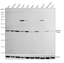
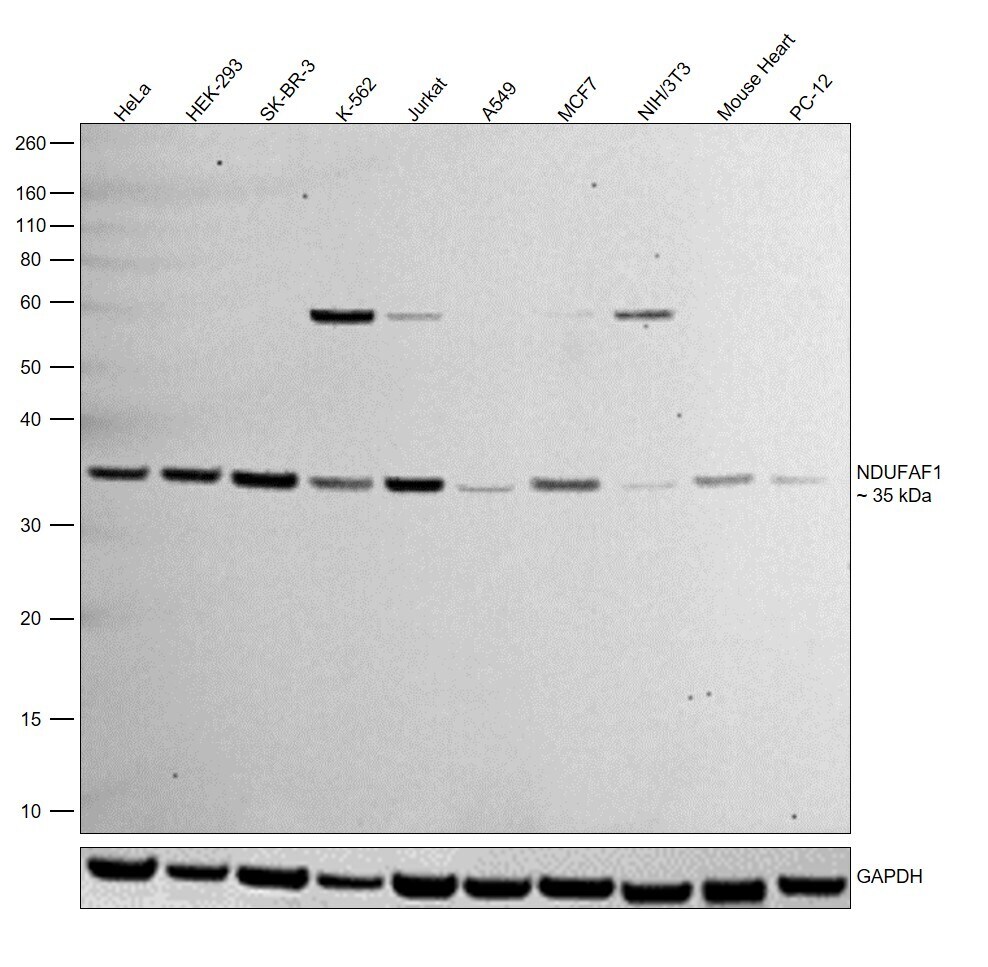
- Western Blot was performed using Anti-NDUFAF1 Polyclonal Antibody (Product # PA5-59051) and a 35 kDa band corresponding to Complex I intermediate-associated protein 30, mitochondrial was observed. Whole cell extracts (30 µg lysate) of HeLa (Lane 1), HEK-293 (Lane 2), SK-BR-3 (Lane 3), K-562 (Lane 4), Jurkat (Lane 5), A549 (Lane 6), MCF7 (Lane 7), NIH/3T3 (Lane 8), tissue extract (30 µg lysate) of Mouse Heart (Lane 9), PC-12 (Lane 10) were electrophoresed using NuPAGE™ 10% Bis-Tris Protein Gel (Product # NP0302BOX). Resolved proteins were then transferred onto a nitrocellulose membrane (Product # IB23001) by iBlot® 2 Dry Blotting System (Product # IB21001). The blot was probed with the primary antibody (0.2 µg/mL) and detected by chemiluminescence with Goat anti-Rabbit IgG (Heavy Chain) Superclonal™ Recombinant Secondary Antibody, HRP (Product # A27036, 1:4000) using the iBright FL 1000 (Product # A32752). Chemiluminescent detection was performed using Novex® ECL Chemiluminescent Substrate Reagent Kit (Product # WP20005).
- Submitted by
- Invitrogen Antibodies (provider)
- Main image
- Experimental details
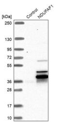
- Western blot analysis of NDUFAF1 in control (vector only transfected HEK293T lysate) and NDUFAF1 over-expression lysate (Co-expressed with a C-terminal myc-DDK tag (~3.1 kDa) in mammalian HEK293T cells). Samples were probed using a NDUFAF1 Polyclonal Antibody (Product # PA5-59051).

Supportive validation
- Submitted by
- Invitrogen Antibodies (provider)
- Main image
- Experimental details
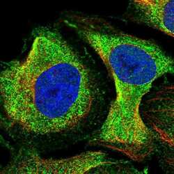
- Immunofluorescent staining of NDUFAF1 in human cell line U-2 OS shows positivity in cytoplasm. Samples were probed using a NDUFAF1 Polyclonal Antibody (Product # PA5-59051).

- Submitted by
- Invitrogen Antibodies (provider)
- Main image

- Experimental details
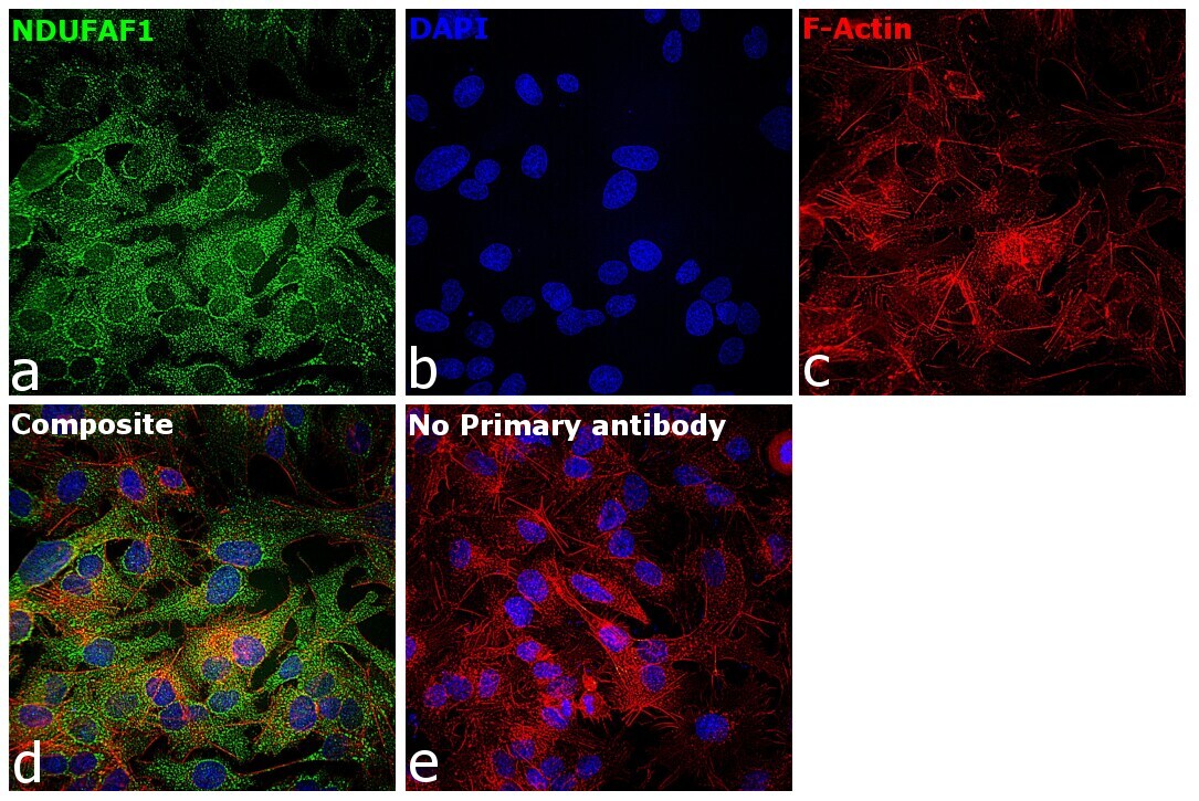
- Immunofluorescence analysis of Complex I intermediate-associated protein 30, mitochondrial was performed using 70% confluent log phase Hep G2 cells. The cells were fixed with 4% paraformaldehyde for 10 minutes, permeabilized with 0.1% Triton™ X-100 for 15 minutes, and blocked with 2% BSA for 45 minutes at room temperature. The cells were labeled with NDUFAF1 Polyclonal Antibody (Product # PA5-59051) at 1:100 in 0.1% BSA, incubated at 4 degree celsius overnight and then labeled with Donkey anti-Rabbit IgG (H+L) Highly Cross-Adsorbed Secondary Antibody, Alexa Fluor Plus 488 (Product # A32790), (1:2000), for 45 minutes at room temperature (Panel a: Green). Nuclei (Panel b:Blue) were stained with Hoechst 33342 (Product # H1399). F-actin (Panel c: Red) was stained with Rhodamine Phalloidin (Product # R415, 1:300). Panel d represents the merged image showing mitochondrial localization. Panel e represents control cells with no primary antibody to assess background. The images were captured at 40X magnification in CellInsight CX7 LZR High-Content Screening (HCS) Platform (Product # CX7C1115LZR).

- Submitted by
- Invitrogen Antibodies (provider)
- Main image

- Experimental details
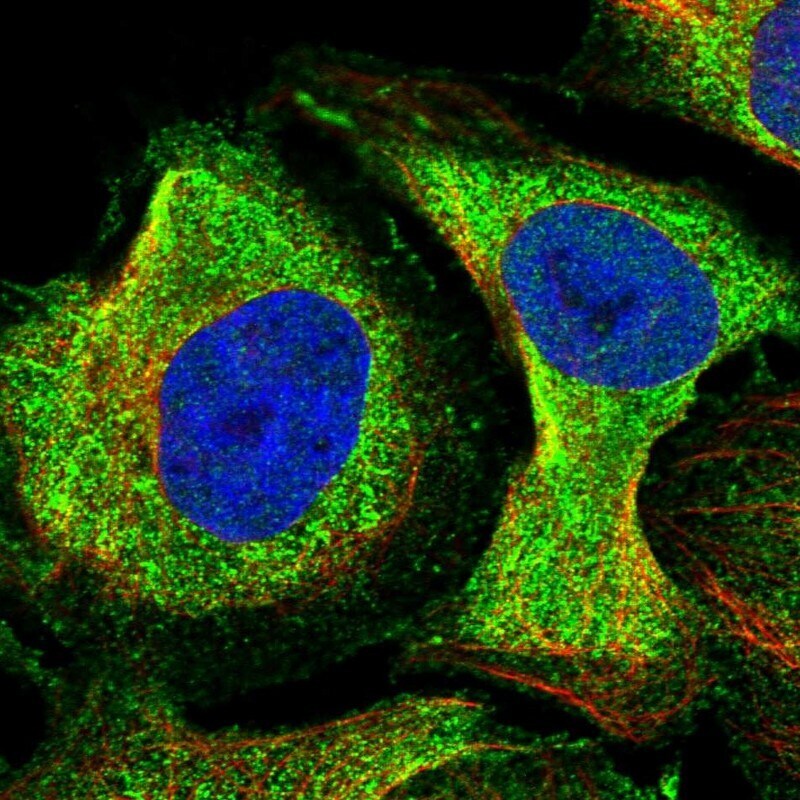
- Immunofluorecent analysis of NDUFAF1 in human cell line U-2 OS using NDUFAF1 Polyclonal Antibody (Product # PA5-59051). Staining shows localization to cytosol.
- Submitted by
- Invitrogen Antibodies (provider)
- Main image

- Experimental details
- Immunofluorescence analysis of Complex I intermediate-associated protein 30, mitochondrial was performed using 70% confluent log phase Hep G2 cells. The cells were fixed with 4% paraformaldehyde for 10 minutes, permeabilized with 0.1% Triton™ X-100 for 15 minutes, and blocked with 2% BSA for 45 minutes at room temperature. The cells were labeled with NDUFAF1 Polyclonal Antibody (Product # PA5-59051) at 1:100 in 0.1% BSA, incubated at 4 degree celsius overnight and then labeled with Donkey anti-Rabbit IgG (H+L) Highly Cross-Adsorbed Secondary Antibody, Alexa Fluor Plus 488 (Product # A32790), (1:2000), for 45 minutes at room temperature (Panel a: Green). Nuclei (Panel b:Blue) were stained with Hoechst 33342 (Product # H1399). F-actin (Panel c: Red) was stained with Rhodamine Phalloidin (Product # R415, 1:300). Panel d represents the merged image showing mitochondrial localization. Panel e represents control cells with no primary antibody to assess background. The images were captured at 40X magnification in CellInsight CX7 LZR High-Content Screening (HCS) Platform (Product # CX7C1115LZR).
Supportive validation
- Submitted by
- Invitrogen Antibodies (provider)
- Main image
- Experimental details
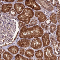
- Immunohistochemical analysis of NDUFAF1 in human kidney using NDUFAF1 Polyclonal Antibody (Product # PA5-59051) shows strong cytoplasmic positivity in cells in tubules.
