 Explore
 Validate
 Learn
Learn Western blot
Antibody data
- Antibody Data
- Antigen structure
- References [1]
- Comments [0]
- Validations
- Western blot [3]
- Immunocytochemistry [3]
- Immunohistochemistry [2]
- Other assay [1]
Submit
Validation data
Reference
Comment
Report error
- Product number
- PA5-31697 - Provider product page

- Provider
- Invitrogen Antibodies
- Product name
- PELO Polyclonal Antibody
- Antibody type
- Polyclonal
- Antigen
- Recombinant full-length protein
- Description
- Recommended positive controls: 293T, HeLa, HepG2, PC-12, Rat2, , Neuro 2A, C8D30, NIH-3T3, Raw264.7, C2C12. Predicted reactivity: Human (99%), Mouse (97%), Rat (97%), Zebrafish (82%), Xenopus laevis (83%), Chicken (95%), Rhesus Monkey (100%), Bovine (98%). Store product as a concentrated solution. Centrifuge briefly prior to opening the vial.
- Reactivity
- Human, Mouse, Rat
- Host
- Rabbit
- Isotype
- IgG
- Vial size
- 100 μL
- Concentration
- 1 mg/mL
- Storage
- Store at 4°C short term. For long term storage, store at -20°C, avoiding freeze/thaw cycles.
Submitted references Coupled protein synthesis and ribosome-guided piRNA processing on mRNAs.
Sun YH, Wang RH, Du K, Zhu J, Zheng J, Xie LH, Pereira AA, Zhang C, Ricci EP, Li XZ
Nature communications 2021 Oct 13;12(1):5970
Nature communications 2021 Oct 13;12(1):5970
No comments: Submit comment
Supportive validation
- Submitted by
- Invitrogen Antibodies (provider)
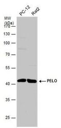
- Main image
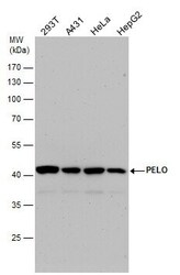
- Experimental details
- PELO Polyclonal Antibody detects PELO protein by western blot analysis. Various whole cell extracts (30 µg) were separated by 10% SDS-PAGE, and the membrane was blotted with PELO Polyclonal Antibody (Product # PA5-31697) diluted by 1:1,000. The HRP-conjugated anti-rabbit IgG antibody was used to detect the primary antibody.
- Submitted by
- Invitrogen Antibodies (provider)
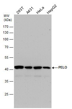
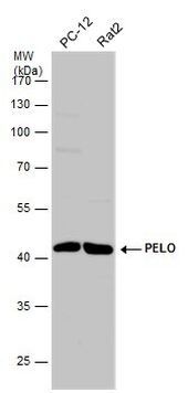
- Main image
- Experimental details
- Western Blot analysis of PELO was performed by separating 30 µg of various whole cell extracts by 10% SDS-PAGE. Proteins were transferred to a membrane and probed with a PELO Polyclonal Antibody (Product # PA5-31697) at a dilution of 1:1000 and a HRP-conjugated anti-rabbit IgG secondary antibody.
- Submitted by
- Invitrogen Antibodies (provider)
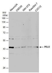
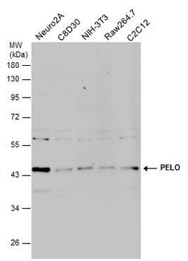
- Main image
- Experimental details
- Western Blot analysis of PELO was performed by separating 30 µg of various whole cell extracts by 10% SDS-PAGE. Proteins were transferred to a membrane and probed with a PELO Polyclonal Antibody (Product # PA5-31697) at a dilution of 1:1000 and a HRP-conjugated anti-rabbit IgG secondary antibody.
Supportive validation
- Submitted by
- Invitrogen Antibodies (provider)
- Main image
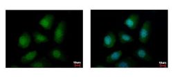
- Experimental details
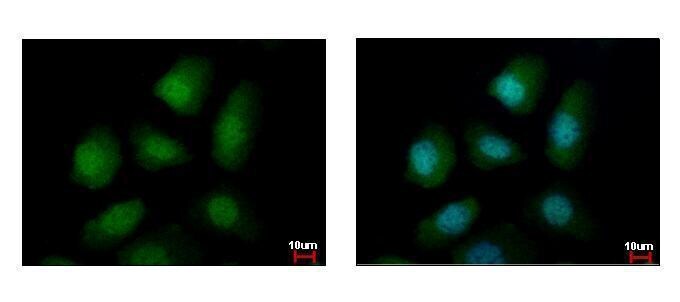
- Immunofluorescent analysis of PELO showing staining in the cytoplasm and nucleus of A431 cells. A431 cells were fixed in 4% paraformaldehyde at RT for 15 min and stained using a PELO polyclonal antibody (Product # PA5-31697) diluted at 1:500. Blue: Hoechst 33342 staining.
- Submitted by
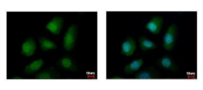
- Invitrogen Antibodies (provider)
- Main image

- Experimental details
- PELO Polyclonal Antibody detects PELO protein at cytoplasm and nucleus by immunofluorescent analysis. Sample: A431 cells were fixed in 4% paraformaldehyde at RT for 15 min. Green: PELO protein stained by PELO Polyclonal Antibody (Product # PA5-31697) diluted at 1:500. Blue: Hoechst 33342 staining.
- Submitted by
- Invitrogen Antibodies (provider)
- Main image

- Experimental details
- PELO Polyclonal Antibody detects PELO protein at cytoplasm and nucleus by immunofluorescent analysis. Sample: A431 cells were fixed in 4% paraformaldehyde at RT for 15 min. Green: PELO protein stained by PELO Polyclonal Antibody (Product # PA5-31697) diluted at 1:500. Blue: Hoechst 33342 staining.

Supportive validation
- Submitted by
- Invitrogen Antibodies (provider)
- Main image
- Experimental details
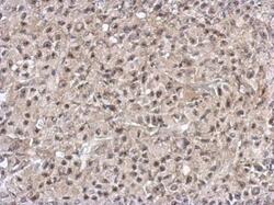
- Immunohistochemical analysis of paraffin-embedded HBL435 xenograft, using PELO (Product # PA5-31697) antibody at 1:500 dilution. Antigen Retrieval: EDTA based buffer, pH 8.0, 15 min.
- Submitted by
- Invitrogen Antibodies (provider)
- Main image
- Experimental details
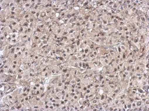
- Immunohistochemical analysis of paraffin-embedded HBL435 xenograft, using PELO (Product # PA5-31697) antibody at 1:500 dilution. Antigen Retrieval: EDTA based buffer, pH 8.0, 15 min.
Supportive validation
- Submitted by
- Invitrogen Antibodies (provider)
- Main image
- Experimental details
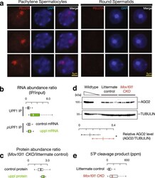
- Fig. 5 3'UTR piRNA biogenesis as a special co-translational mRNA decay pathway. a Immunolabeling of squashed pachytene spermatocytes (left) and round spermatids (right). Three examples of each stage were shown using anti-PELOTA, DAPI, and merged. Experiments were repeated at least three times independently with similar results. Scale bar, 5 mum. b Boxplots of the ratios of RNA abundance in UPF1 IP (upper) and phosphorylated UPF1 IP mutant (lower) versus their corresponding input in adult wild-type testes. Control mRNA n = 43, uppl mRNA n = 30. c Boxplots of the changes of protein abundance coded per gene in Mov10l1 mutants (sample size n = 5 independent biological replicates) compared to littermate controls in adult testes. Control mRNA n = 43, uppl mRNA n = 30. d AGO2 western blot (top) and TUBULIN western blot (bottom) of adult testis lysates from serial dilutions of adult wild-type lysates (left) and four biological replicates of Mov10l1 CKO/ Neurog3-cre (middle) and Mov10l1 CKO/ (right). Abundance of AGO2 and TUBULIN was quantified using ImageJ and then fitted to the standard curve of the wild-type testis lysates. Relative abundance of AGO2 were calculated as the abundance of AGO2 normalized by the abundance of TUBULIN (sample size n = 4 independent biological samples). Data are mean +- standard deviation; * p = 0.048, one-side Student's t test. Each data point was overlaid as dot plots. e Boxplots of the 5'P decay intermediates per mRNA in adult wild-type testes. AGO2 ta
