 Explore
 Validate
 Learn
Learn Western blot
 Immunocytochemistry
Antibody data
- Antibody Data
- Antigen structure
- References [2]
- Comments [0]
- Validations
- Immunocytochemistry [1]
Submit
Validation data
Reference
Comment
Report error
- Product number
- HPA040454 - Provider product page

- Provider
- Atlas Antibodies
- Proper citation
- Atlas Antibodies Cat#HPA040454, RRID:AB_10672897
- Product name
- Anti-ITPKA
- Antibody type
- Polyclonal
- Description
- Polyclonal Antibody against Human ITPKA, Gene description: inositol-trisphosphate 3-kinase A, Alternative Gene Names: IP3-3KA, IP3KA, Validated applications: ICC, WB, Uniprot ID: P23677, Storage: Store at +4°C for short term storage. Long time storage is recommended at -20°C.
- Reactivity
- Human
- Host
- Rabbit
- Conjugate
- Unconjugated
- Isotype
- IgG
- Vial size
- 100 µl
- Concentration
- 0.1 mg/ml
- Storage
- Store at +4°C for short term storage. Long time storage is recommended at -20°C.
- Handling
- The antibody solution should be gently mixed before use.
Submitted references ITPKA phosphorylates PYCR1 and promotes the progression of glioma
ITPKA induces cell senescence, inhibits ovarian cancer tumorigenesis and can be downregulated by miR-203
Luo X, Chen T, Deng J, Liu Z, Bi C, Lan S
Heliyon 2024;10(15):e35303
Heliyon 2024;10(15):e35303
ITPKA induces cell senescence, inhibits ovarian cancer tumorigenesis and can be downregulated by miR-203
Shaosheng W, Shaochuang W, Lichun F, Na X, Xiaohong Z
Aging 2021;13(8):11822-11832
Aging 2021;13(8):11822-11832
No comments: Submit comment
Supportive validation
- Submitted by
- Atlas Antibodies (provider)
- Main image
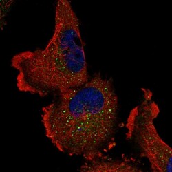
- Experimental details
- Immunofluorescent staining of human cell line U-251 MG shows localization to vesicles.
- Sample type
- Human
