Explore
 Validate
 Learn
Learn Western blot
 Immunocytochemistry
Antibody data
- Antibody Data
- Antigen structure
- References [1]
- Comments [0]
- Validations
- Immunocytochemistry [2]
- Immunohistochemistry [1]
- Flow cytometry [3]
- Other assay [2]
Submit
Validation data
Reference
Comment
Report error
- Product number
- PA5-47677 - Provider product page

- Provider
- Invitrogen Antibodies
- Product name
- CLEC14A Polyclonal Antibody
- Antibody type
- Polyclonal
- Antigen
- Recombinant full-length protein
- Description
- In direct ELISAs and Western blots, less than 1% cross-reactivity with recombinant human (rh) CLEC1, rhCLEC2, rhCLEC3B, and rhCLEC10A is observed. Reconstitute at 0.2 mg/mL in sterile PBS.
- Reactivity
- Human
- Host
- Sheep
- Isotype
- IgG
- Vial size
- 100 μg
- Concentration
- 0.2 mg/mL
- Storage
- -20°C, Avoid Freeze/Thaw Cycles
Submitted references Human muscle-derived CLEC14A-positive cells regenerate muscle independent of PAX7.
Marg A, Escobar H, Karaiskos N, Grunwald SA, Metzler E, Kieshauer J, Sauer S, Pasemann D, Malfatti E, Mompoint D, Quijano-Roy S, Boltengagen A, Schneider J, Schülke M, Kunz S, Carlier R, Birchmeier C, Amthor H, Spuler A, Kocks C, Rajewsky N, Spuler S
Nature communications 2019 Dec 18;10(1):5776
Nature communications 2019 Dec 18;10(1):5776
No comments: Submit comment
Supportive validation
- Submitted by
- Invitrogen Antibodies (provider)
- Main image

- Experimental details
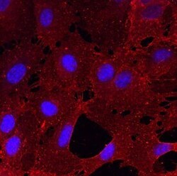
- Immunocytochemistry analysis of CLEC14A in immersion fixed HUVEC human umbilical vein endothelial cells. Samples were incubated in CLEC14A polyclonal antibody (Product # PA5-47677) using a dilution of 10 µg/mL for 3 hours at room temperature followed by NorthernLights™ 557-conjugated Anti-Sheep IgG Secondary Antibody (red) and counterstained with DAPI (blue). Specific staining was localized to cell surfaces and cytoplasm.

- Submitted by
- Invitrogen Antibodies (provider)
- Main image
- Experimental details
- Immunocytochemistry analysis of CLEC14A in immersion fixed HUVEC human umbilical vein endothelial cells. Samples were incubated in CLEC14A polyclonal antibody (Product # PA5-47677) using a dilution of 10 µg/mL for 3 hours at room temperature followed by NorthernLights™ 557-conjugated Anti-Sheep IgG Secondary Antibody (red) and counterstained with DAPI (blue). Specific staining was localized to cell surfaces and cytoplasm.

Supportive validation
- Submitted by
- Invitrogen Antibodies (provider)
- Main image
- Experimental details
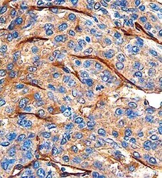
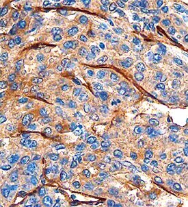
- Immunohistochemical analysis of CLEC14A in formalin fixed paraffin-embedded sections of human breast cancer tissue. Samples were incubated in CLEC14A polyclonal antibody (Product # PA5-47677) using a dilution of 1.7 µg/mL overnight at 4 °C. Before incubation with the primary antibody, tissue was subjected to heat-induced epitope retrieval using Antigen Retrieval Reagent-Universal . Tissue was stained using the Anti-Sheep HRP-DAB Cell & Tissue Staining Kit (brown) and counterstained with hematoxylin (blue). Specific staining was localized to endothelial cells.
Supportive validation
- Submitted by
- Invitrogen Antibodies (provider)
- Main image
- Experimental details
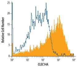
- Flow cytometric analysis of HUVEC human umbilical vein endothelial cells were stained with Sheep Anti-human CLEC14A Antigen Affinity-purified Polyclonal Antibody (Product # PA5-47677, filled histogram) or isotype control antibody (open histogram), followed by Phycoerythrin-conjugated Anti-Sheep IgG Secondary Antibody.

- Submitted by
- Invitrogen Antibodies (provider)
- Main image
- Experimental details
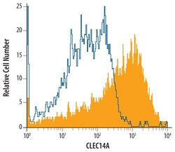
- Flow cytometry of CLEC14A of HUVEC human umbilical vein endothelial cells. Samples were incubated in CLEC14A polyclonal antibody (Product # PA5-47677) or isotype control antibody followed by Phycoerythrin-conjugated Anti-Sheep IgG Secondary Antibody.

- Submitted by
- Invitrogen Antibodies (provider)
- Main image

- Experimental details
- Flow cytometry of CLEC14A of HUVEC human umbilical vein endothelial cells. Samples were incubated in CLEC14A polyclonal antibody (Product # PA5-47677) or isotype control antibody followed by Phycoerythrin-conjugated Anti-Sheep IgG Secondary Antibody.

Supportive validation
- Submitted by
- Invitrogen Antibodies (provider)
- Main image
- Experimental details
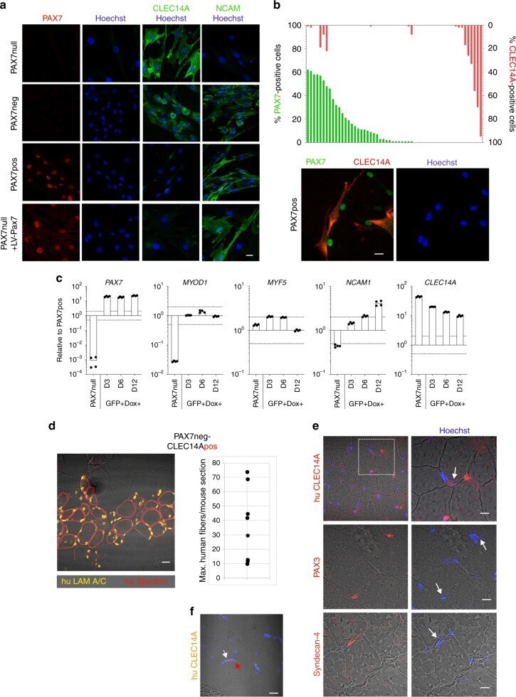
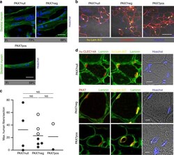
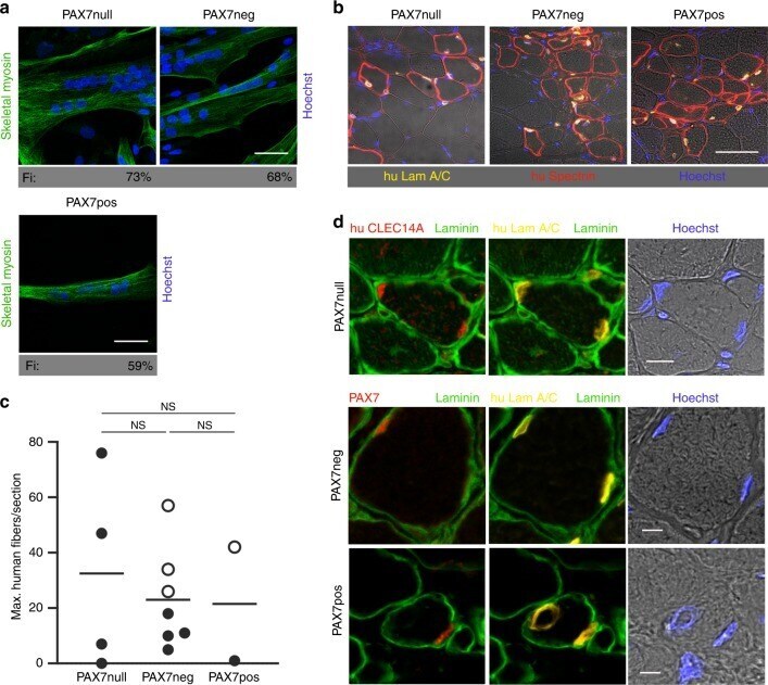
- Fig. 3 PAX7-negative cell populations can generate muscle. a PAX7, PAX7neg, and PAX7pos cells, used in transplantation experiments shown in b , indicate the same differentiation potential (skeletal myosin staining, Fi: Fusion index, Scale bars: 50 um). b TA muscle sections from NOG mice demonstrate that 3 weeks after transplantation the PAX7, PAX7neg, and PAX7pos human cell types contributed to muscle regeneration. Human anti-lamin A/C antibody (yellow) detects only human nuclei and anti-human spectrin antibody is specific for fibers of human origin (red). Nuclei in blue (Hoechst). Scale bar: 50 um. c Quantification of experiments shown in b . Each dot represents one mouse grafted with the cells indicated below ( n = 4 for PAX7 and n = 7 for PAX7neg). Shown are the values for the section with the highest number of human fibers for each mouse and the means. Only human muscle fibers with a diameter > 10 um were counted. Mice with > 20 human fibers/section were additionally analyzed for PAX7 expression. The presence of PAX7-positive satellite cells is indicated by an open circle. The statistical differences between the groups were analyzed by two-tailed t tests, P > 0.05 = NS (not significant). d Repopulation of satellite cell niche after transplantation: In PAX7 transplants, the satellite cell niche is populated by CLEC14A-positive cells (red, upper row). After transplantation of PAX7neg- and PAX7pos cells, the satellite cell niche is populated by PAX7-positive
- Submitted by
- Invitrogen Antibodies (provider)
- Main image
- Experimental details
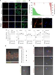
- Fig. 4 CLEC14A is a marker for a human myogenic cell in satellite cell position. a CLEC14A- staining of PAX7, PAX7neg, PAX7pos cells. PAX7 cells are strongly positive for CLEC14A. Some PAX7neg cell populations from dornors with intact PAX7 gene are also CLEC14A-positive. Reconstitution of PAX7 cells with a lentiviral PAX7 vector changed the expression profile of CLEC14A and NCAM (bottom line). Red, PAX7; green, CLEC14A; blue, Hoechst. Bar 20 um. b PAX7 and CLEC14A expression in PAX7neg cell colonies were mutual exclusive. Fifty-six cell populations from donors with intact PAX7 genes were analyzed for expression of PAX7 (green) and CLEC14A (red). Cell colonies are either positive for PAX7 or for CLEC14A. Some colonies contained both, cells that were either PAX7 or CLEC14A-positive. Staining for PAX7 and CLEC14A in ""mixed"" cell populations depicted mutually exclusive expression. Scale bar: 20 um. c Gene expression in PAX7 cells selected by FACS-sorting (see also Supplementary Fig. 9 ) after lentiviral PAX7 transduction determined by qPCR 3, 6, and 12 days after DOX induction. All data were normalized to two reference genes and relativized to the mean of PAX7pos cell colonies. The dotted line at twofold and 0.5-fold represents the threshold for differential expression. d TA muscle section from a NOG mouse 3 weeks after transplantation of CLEC14Apos cells shows newly built human muscle fibers. Quantification reveals similar results as in Fig. 3b . Quantification