 Explore
 Validate
 Learn
Learn Western blot
Antibody data
- Antibody Data
- Antigen structure
- References [1]
- Comments [0]
- Validations
- Western blot [3]
- Immunocytochemistry [3]
- Immunohistochemistry [3]
Submit
Validation data
Reference
Comment
Report error
- Product number
- PA5-28984 - Provider product page

- Provider
- Invitrogen Antibodies
- Product name
- SEC23A Polyclonal Antibody
- Antibody type
- Polyclonal
- Antigen
- Recombinant full-length protein
- Description
- Recommended positive controls: 293T, A431, H1299, HeLa, HepG2, Molt-4, Raji. Predicted reactivity: Mouse (98%), Rat (98%), Zebrafish (87%), Rhesus Monkey (99%), Bovine (99%). Store product as a concentrated solution. Centrifuge briefly prior to opening the vial.
- Reactivity
- Human, Mouse
- Host
- Rabbit
- Isotype
- IgG
- Vial size
- 100 μL
- Concentration
- 1 mg/mL
- Storage
- Store at 4°C short term. For long term storage, store at -20°C, avoiding freeze/thaw cycles.
Submitted references ICAM1 initiates CTC cluster formation and trans-endothelial migration in lung metastasis of breast cancer.
Taftaf R, Liu X, Singh S, Jia Y, Dashzeveg NK, Hoffmann AD, El-Shennawy L, Ramos EK, Adorno-Cruz V, Schuster EJ, Scholten D, Patel D, Zhang Y, Davis AA, Reduzzi C, Cao Y, D'Amico P, Shen Y, Cristofanilli M, Muller WA, Varadan V, Liu H
Nature communications 2021 Aug 11;12(1):4867
Nature communications 2021 Aug 11;12(1):4867
No comments: Submit comment
Supportive validation
- Submitted by
- Invitrogen Antibodies (provider)
- Main image

- Experimental details
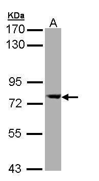
- Western Blot using SEC23A Polyclonal Antibody (Product # PA5-28984). Sample (30 µg of whole cell lysate). A: Molt-4. 7.5% SDS PAGE. SEC23A Polyclonal Antibody (Product # PA5-28984) diluted at 1:10,000. The HRP-conjugated anti-rabbit IgG antibody was used to detect the primary antibody.
- Submitted by
- Invitrogen Antibodies (provider)
- Main image
- Experimental details
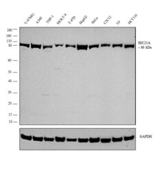
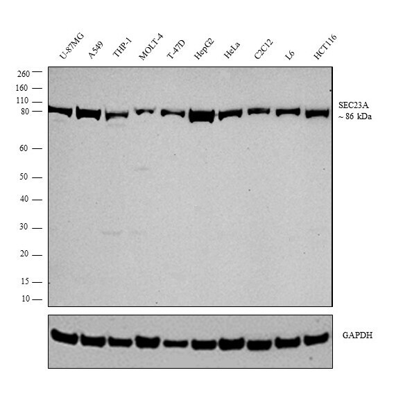
- Western blot analysis was performed on whole cell extract (30 µg lysate) of U-87 mg (Lane 1), A549 (Lane 2), THP-1 (Lane 3), MOLT-4 (Lane 4), T-47D (Lane 5), HepG2 (Lane 6), HeLa (Lane 7), C2C12 (Lane 8), L6 (Lane 9) and HCT116 (Lane 10). The blot was probed with Anti-SEC23A Polyclonal Antibody (Product # PA5-28984, 1:10,000 dilution) and detected by chemiluminescence using Goat anti-Rabbit IgG (Heavy Chain) Superclonal™ Secondary Antibody, HRP conjugate (Product # A27036, 0.25 µg/mL, 1:4,000 dilution). A 86 kDa band corresponding to SEC23A was observed across the cell lines tested.
- Submitted by
- Invitrogen Antibodies (provider)
- Main image

- Experimental details
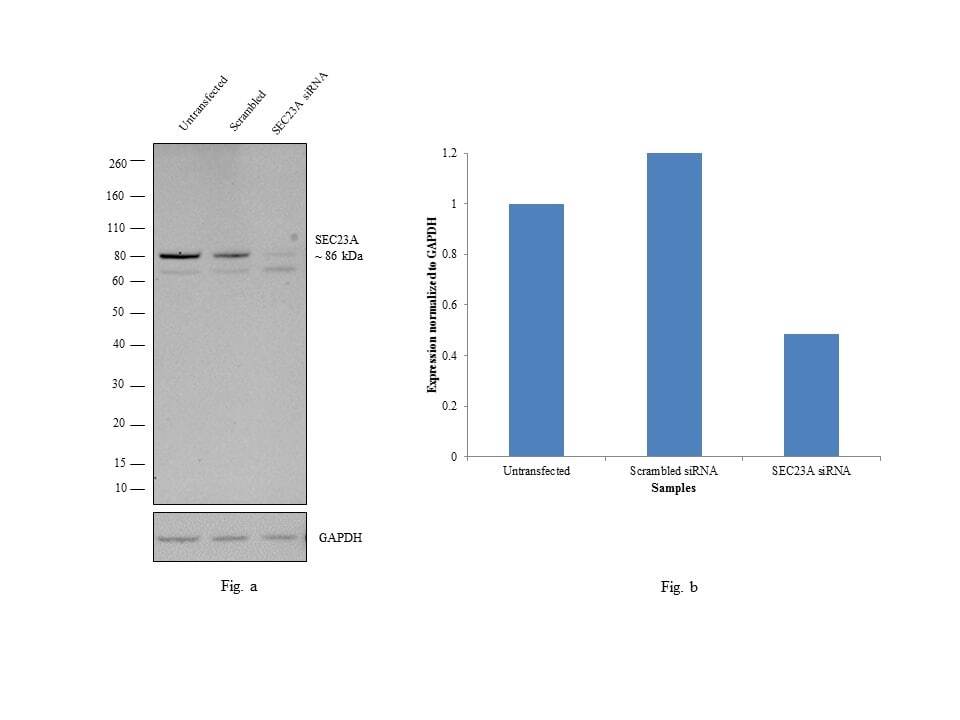
- Knockdown of SEC23A was achieved by transfecting A549 cells with SEC23A specific siRNAs (Silencer® select Product # s20538, s20539). Western blot analysis (Fig. a) was performed using whole cell extracts from the SEC23A knockdown cells (lane 3), non-specific scrambled siRNA transfected cells (lane 2) and untransfected cells (lane 1). The blot was probed with SEC23A Polyclonal Antibody (Product # PA5-28984, 1:10,000 dilution) and Goat anti-Rabbit IgG (Heavy Chain) Superclonal™ Secondary Antibody, HRP conjugate (Product # A27036, 0.25 µg/mL, 1:4,000 dilution). Densitometric analysis of this western blot is shown in histogram (Fig. b). Decrease in signal upon siRNA mediated knock down confirms that antibody is specific to SEC23A.
Supportive validation
- Submitted by
- Invitrogen Antibodies (provider)
- Main image
- Experimental details
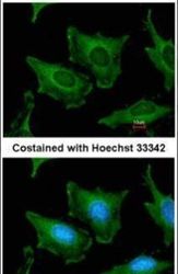
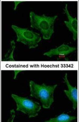
- Immunofluorescent analysis of SEC23A in methanol-fixed HeLa cells using a SEC23A polyclonal antibody (Product # PA5-28984) at a 1:200 dilution.
- Submitted by
- Invitrogen Antibodies (provider)
- Main image

- Experimental details
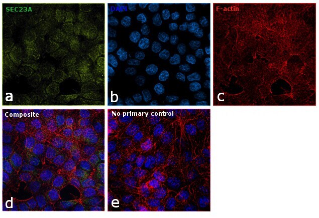
- Immunofluorescence analysis of SEC23A was performed using 70% confluent log phase HCT 116 cells. The cells were fixed with 4% paraformaldehyde for 10 minutes, permeabilized with 0.1% Triton™ X-100 for 15 minutes, and blocked with 1% BSA for 1 hour at room temperature. The cells were labeled with SEC23A Polyclonal Antibody (Product # PA5-28984) at 1:200 dilution in 0.1% BSA, incubated at 4 degree Celsius overnight and then labeled with Goat anti-Rabbit IgG (H+L) Superclonal™ Secondary Antibody, Alexa Fluor® 488 conjugate (Product # A27034) at a dilution of 1:2000 for 45 minutes at room temperature (Panel a: green). Nuclei (Panel b: blue) were stained with SlowFade® Gold Antifade Mountant with DAPI (Product # S36938). F-actin (Panel c: red) was stained with Rhodamine Phalloidin (Product # R415, 1:300). Panel d represents the merged image showing cytoplasmic localization. Panel e represents control cells with no primary antibody to assess background. The images were captured at 60X magnification.
- Submitted by
- Invitrogen Antibodies (provider)
- Main image
- Experimental details
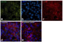
- Immunofluorescence analysis of SEC23A was performed using 70% confluent log phase HCT 116 cells. The cells were fixed with 4% paraformaldehyde for 10 minutes, permeabilized with 0.1% Triton™ X-100 for 15 minutes, and blocked with 1% BSA for 1 hour at room temperature. The cells were labeled with SEC23A Polyclonal Antibody (Product # PA5-28984) at 1:200 dilution in 0.1% BSA, incubated at 4 degree Celsius overnight and then labeled with Goat anti-Rabbit IgG (Heavy Chain) Superclonal™ Secondary Antibody, Alexa Fluor® 488 conjugate (Product # A27034) at a dilution of 1:2000 for 45 minutes at room temperature (Panel a: green). Nuclei (Panel b: blue) were stained with SlowFade® Gold Antifade Mountant with DAPI (Product # S36938). F-actin (Panel c: red) was stained with Rhodamine Phalloidin (Product # R415, 1:300). Panel d represents the merged image showing cytoplasmic localization. Panel e represents control cells with no primary antibody to assess background. The images were captured at 60X magnification.

Supportive validation
- Submitted by
- Invitrogen Antibodies (provider)
- Main image
- Experimental details
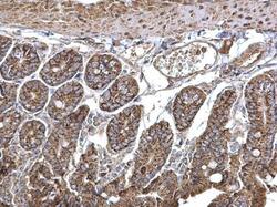
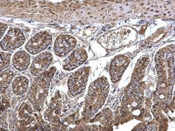
- SEC23A Polyclonal Antibody detects SEC23A protein at cytosol on mouse intestine by immunohistochemical analysis. Sample: Paraffin-embedded mouse intestine. SEC23A Polyclonal Antibody (Product # PA5-28984) dilution: 1:500. Antigen Retrieval: EDTA based buffer, pH 8.0, 15 min.
- Submitted by
- Invitrogen Antibodies (provider)
- Main image
- Experimental details
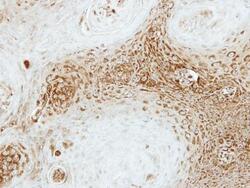
- Immunohistochemical analysis of paraffin-embedded Cal27 xenograft, using SEC23A (Product # PA5-28984) antibody at 1:500 dilution. Antigen Retrieval: EDTA based buffer, pH 8.0, 15 min.

- Submitted by
- Invitrogen Antibodies (provider)
- Main image
- Experimental details
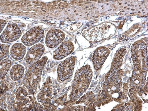
- SEC23A Polyclonal Antibody detects SEC23A protein at cytosol on mouse intestine by immunohistochemical analysis. Sample: Paraffin-embedded mouse intestine. SEC23A Polyclonal Antibody (Product # PA5-28984) dilution: 1:500. Antigen Retrieval: EDTA based buffer, pH 8.0, 15 min.
