 Explore
 Validate
 Learn
Learn Western blot
 Immunocytochemistry
Antibody data
- Antibody Data
- Antigen structure
- References [9]
- Comments [0]
- Validations
- Immunocytochemistry [1]
- Immunohistochemistry [1]
Submit
Validation data
Reference
Comment
Report error
- Product number
- HPA012145 - Provider product page

- Provider
- Atlas Antibodies
- Proper citation
- Atlas Antibodies Cat#HPA012145, RRID:AB_1844494
- Product name
- Anti-ACBD5
- Antibody type
- Polyclonal
- Description
- Polyclonal Antibody against Human ACBD5, Gene description: acyl-CoA binding domain containing 5, Alternative Gene Names: DKFZp434A2417, KIAA1996, Validated applications: IHC, WB, ICC, Uniprot ID: Q5T8D3, Storage: Store at +4°C for short term storage. Long time storage is recommended at -20°C.
- Reactivity
- Human
- Host
- Rabbit
- Conjugate
- Unconjugated
- Isotype
- IgG
- Vial size
- 100 µl
- Storage
- Store at +4°C for short term storage. Long time storage is recommended at -20°C.
- Handling
- The antibody solution should be gently mixed before use.
Submitted references Studying the topology of peroxisomal acyl-CoA synthetases using self-assembling split sfGFP
The subset of peroxisomal tail-anchored proteins do not reach peroxisomes via ER, instead mitochondria can be involved.
Restructured membrane contacts rewire organelles for human cytomegalovirus infection
Autophagy Inhibitors Do Not Restore Peroxisomal Functions in Cells With the Most Common Peroxisome Biogenesis Defect.
Quantitative Proteomics and Differential Protein Abundance Analysis after the Depletion of PEX3 from Human Cells Identifies Additional Aspects of Protein Targeting to the ER
Prostate Cancer Proliferation Is Affected by the Subcellular Localization of MCT2 and Accompanied by Significant Peroxisomal Alterations
Spastin tethers lipid droplets to peroxisomes and directs fatty acid trafficking through ESCRT-III
Fluorescent Tools to Analyze Peroxisome–Endoplasmic Reticulum Interactions in Mammalian Cells
Intracellular redistribution of neuronal peroxisomes in response to ACBD5 expression
Chornyi S, Koster J, IJlst L, Waterham H
Histochemistry and Cell Biology 2024;161(2):133-144
Histochemistry and Cell Biology 2024;161(2):133-144
The subset of peroxisomal tail-anchored proteins do not reach peroxisomes via ER, instead mitochondria can be involved.
Somborac T, Lutfullahoglu Bal G, Fatima K, Vihinen H, Paatero A, Jokitalo E, Paavilainen VO, Konovalova S
PloS one 2023;18(12):e0295047
PloS one 2023;18(12):e0295047
Restructured membrane contacts rewire organelles for human cytomegalovirus infection
Cook K, Tsopurashvili E, Needham J, Thompson S, Cristea I
Nature Communications 2022;13(1)
Nature Communications 2022;13(1)
Autophagy Inhibitors Do Not Restore Peroxisomal Functions in Cells With the Most Common Peroxisome Biogenesis Defect.
Klouwer FCC, Falkenberg KD, Ofman R, Koster J, van Gent D, Ferdinandusse S, Wanders RJA, Waterham HR
Frontiers in cell and developmental biology 2021;9:661298
Frontiers in cell and developmental biology 2021;9:661298
Quantitative Proteomics and Differential Protein Abundance Analysis after the Depletion of PEX3 from Human Cells Identifies Additional Aspects of Protein Targeting to the ER
Zimmermann R, Lang S, Lerner M, Förster F, Nguyen D, Helms V, Schrul B
International Journal of Molecular Sciences 2021;22(23):13028
International Journal of Molecular Sciences 2021;22(23):13028
Prostate Cancer Proliferation Is Affected by the Subcellular Localization of MCT2 and Accompanied by Significant Peroxisomal Alterations
Valença I, Ferreira A, Correia M, Kühl S, van Roermund C, Waterham H, Máximo V, Islinger M, Ribeiro D
Cancers 2020;12(11):3152
Cancers 2020;12(11):3152
Spastin tethers lipid droplets to peroxisomes and directs fatty acid trafficking through ESCRT-III
Chang C, Weigel A, Ioannou M, Pasolli H, Xu C, Peale D, Shtengel G, Freeman M, Hess H, Blackstone C, Lippincott-Schwartz J
Journal of Cell Biology 2019;218(8):2583-2599
Journal of Cell Biology 2019;218(8):2583-2599
Fluorescent Tools to Analyze Peroxisome–Endoplasmic Reticulum Interactions in Mammalian Cells
Bishop A, Kamoshita M, Passmore J, Hacker C, Schrader T, Waterham H, Costello J, Schrader M
Contact 2019;2
Contact 2019;2
Intracellular redistribution of neuronal peroxisomes in response to ACBD5 expression
Zalckvar E, Wang Y, Metz J, Costello J, Passmore J, Schrader M, Schultz C, Islinger M
PLOS ONE 2018;13(12):e0209507
PLOS ONE 2018;13(12):e0209507
No comments: Submit comment
Supportive validation
- Submitted by
- Atlas Antibodies (provider)
- Main image
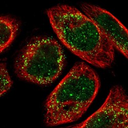
- Experimental details
- Immunofluorescent staining of human cell line A-431 shows localization to nucleus & peroxisomes.
- Sample type
- Human
Supportive validation
- Submitted by
- Atlas Antibodies (provider)
- Enhanced method
- Orthogonal validation
- Main image
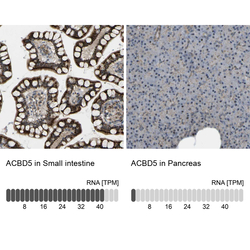
- Experimental details
- Immunohistochemistry analysis in human small intestine and pancreas tissues using HPA012145 antibody. Corresponding ACBD5 RNA-seq data are presented for the same tissues.
- Sample type
- Human
- Protocol
- Protocol
