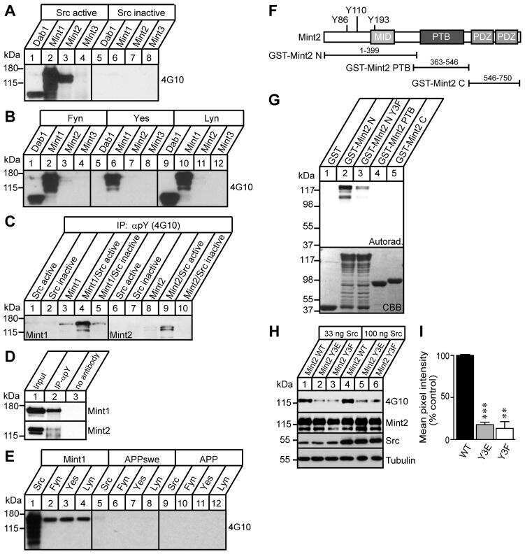
Explore
 Validate
 Learn
Learn Western blot
Antibody data
- Antibody Data
- Antigen structure
- References [8]
- Comments [0]
- Validations
- Western blot [2]
- Immunocytochemistry [2]
- Flow cytometry [1]
- Other assay [1]
Submit
Validation data
Reference
Comment
Report error
- Product number
- PA1-072 - Provider product page

- Provider
- Invitrogen Antibodies
- Product name
- MINT3 Polyclonal Antibody
- Antibody type
- Polyclonal
- Antigen
- Synthetic peptide
- Description
- PA1-072 detects munc-18 interacting protein 3 (Mint3) from mouse cells and rat brain samples. PA1-072 has been successfully used in Western blot procedures. By Western blot, this antibody detects an ~86 kDa protein representing Mint3 from AtT20 cell extract and rat brain samples. The PA1-072 immunogen is a synthetic peptide corresponding to residues M(1) E F L P E P Q H P P G P P T M D L E(19) of rat Mint3. The PA1-072 immunizing peptide (Cat. # PEP-135) is available for use in neutralization and control experiments.
- Reactivity
- Human, Mouse, Rat
- Host
- Rabbit
- Isotype
- IgG
- Vial size
- 100 μg
- Concentration
- 1 mg/mL
- Storage
- -20°C, Avoid Freeze/Thaw Cycles
Submitted references Downregulation of microRNA-448 improves isoflurane-induced learning and memory impairment in rats.
Mint proteins are required for synaptic activity-dependent amyloid precursor protein (APP) trafficking and amyloid β generation.
Intracellular amyloid precursor protein sorting and amyloid-β secretion are regulated by Src-mediated phosphorylation of Mint2.
Regulation of APP-dependent transcription complexes by Mint/X11s: differential functions of Mint isoforms.
Regulation of APP-dependent transcription complexes by Mint/X11s: differential functions of Mint isoforms.
Amyloid precursor protein associates independently and collaboratively with PTB and PDZ domains of mint on vesicles and at cell membrane.
Neuronal expression of mint1 and mint2, novel multimodular proteins, in adult murine brain.
Neuronal expression of mint1 and mint2, novel multimodular proteins, in adult murine brain.
Wu Q, Dai Q, Jiang L, Wang Y, Yang T, Miao J, Wang J, Han Y
Molecular medicine reports 2017 Aug;16(2):1578-1583
Molecular medicine reports 2017 Aug;16(2):1578-1583
Mint proteins are required for synaptic activity-dependent amyloid precursor protein (APP) trafficking and amyloid β generation.
Sullivan SE, Dillon GM, Sullivan JM, Ho A
The Journal of biological chemistry 2014 May 30;289(22):15374-83
The Journal of biological chemistry 2014 May 30;289(22):15374-83
Intracellular amyloid precursor protein sorting and amyloid-β secretion are regulated by Src-mediated phosphorylation of Mint2.
Chaufty J, Sullivan SE, Ho A
The Journal of neuroscience : the official journal of the Society for Neuroscience 2012 Jul 11;32(28):9613-25
The Journal of neuroscience : the official journal of the Society for Neuroscience 2012 Jul 11;32(28):9613-25
Regulation of APP-dependent transcription complexes by Mint/X11s: differential functions of Mint isoforms.
Biederer T, Cao X, Südhof TC, Liu X
The Journal of neuroscience : the official journal of the Society for Neuroscience 2002 Sep 1;22(17):7340-51
The Journal of neuroscience : the official journal of the Society for Neuroscience 2002 Sep 1;22(17):7340-51
Amyloid precursor protein associates independently and collaboratively with PTB and PDZ domains of mint on vesicles and at cell membrane.
Okamoto M, Nakajima Y, Matsuyama T, Sugita M
Neuroscience 2001;104(3):653-65
Neuroscience 2001;104(3):653-65
Neuronal expression of mint1 and mint2, novel multimodular proteins, in adult murine brain.
Nakajima Y, Okamoto M, Nishimura H, Obata K, Kitano H, Sugita M, Matsuyama T
Brain research. Molecular brain research 2001 Aug 15;92(1-2):27-42
Brain research. Molecular brain research 2001 Aug 15;92(1-2):27-42
No comments: Submit comment
Supportive validation
- Submitted by
- Invitrogen Antibodies (provider)
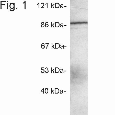
- Main image

- Experimental details
- Western blot detection of Mint3 from AtT20 cell extract using Product # PA1-072.
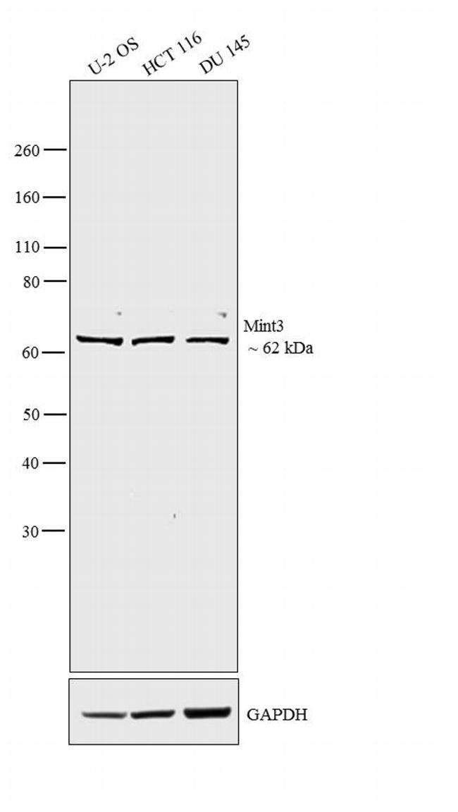
- Submitted by
- Invitrogen Antibodies (provider)
- Main image
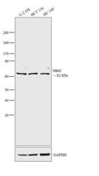
- Experimental details
- Western blot analysis was performed on whole cell extracts (30 µg lysate) of U-2 OS (Lane 1), HCT 116 (Lane 2) and DU 145 (Lane 3). The blots were probed with Anti-Mint3 Rabbit Polyclonal Antibody (Product # PA1-072, 2 µg/mL) and detected by chemiluminescence using Goat anti-Rabbit IgG (Heavy Chain) Superclonal™ Secondary Antibody, HRP conjugate (Product # A27036, 0.25 µg/mL, 1:4000 dilution). A 62 kDa band corresponding to Mint3 was observed across the cell lines tested. Known quantity of protein samples were electrophoresed using Novex® NuPAGE® 4-12 % Bis-Tris gel (Product # NP0321BOX), XCell SureLock™ Electrophoresis System (Product # EI0002) and Novex® Sharp Pre-Stained Protein Standard (Product # LC5800). Resolved proteins were then transferred onto a nitrocellulose membrane with iBlot® 2 Dry Blotting System (Product # IB21001). The membrane was probed with the relevant primary and secondary Antibody following blocking with 5 % skimmed milk. Chemiluminescent detection was performed using Pierce™ ECL Western Blotting Substrate (Product # 32106).
Supportive validation
- Submitted by
- Invitrogen Antibodies (provider)
- Main image
- Experimental details
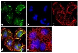
- Immunofluorescence analysis of Mint3 was performed using 90% confluent log phase Neuro-2a cells. The cells were fixed with 4% paraformaldehyde for 10 minutes, permeabilized with 0.1% Triton™ X-100 for 10 minutes, and blocked with 1% BSA for 1 hour at room temperature. The cells were labeled with Mint3 Rabbit Polyclonal Antibody (Product # PA1-072) at 2µg/mL in 0.1% BSA and incubated for 3 hours at room temperature and then labeled with Goat anti-Rabbit IgG (H+L) Superclonal™ Secondary Antibody, Alexa Fluor® 488 conjugate (Product # A27034) at a dilution of 1:2000 for 45 minutes at room temperature (Panel a: green). Nuclei (Panel b: blue) were stained with SlowFade® Gold Antifade Mountant with DAPI (Product # S36938). F-actin (Panel c: red) was stained with Alexa Fluor® 555 Rhodamine Phalloidin (Product # R415, 1:300). Panel d represents the merged image showing membranous localization. Panel e shows the no primary antibody control. The images were captured at 60X magnification.
- Submitted by
- Invitrogen Antibodies (provider)
- Main image

- Experimental details
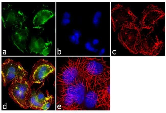
- Immunofluorescence analysis of Mint3 was performed using 90% confluent log phase Neuro-2a cells. The cells were fixed with 4% paraformaldehyde for 10 minutes, permeabilized with 0.1% Triton™ X-100 for 10 minutes, and blocked with 1% BSA for 1 hour at room temperature. The cells were labeled with Mint3 Rabbit Polyclonal Antibody (Product # PA1-072) at 2µg/mL in 0.1% BSA and incubated for 3 hours at room temperature and then labeled with Goat anti-Rabbit IgG (Heavy Chain) Superclonal™ Secondary Antibody, Alexa Fluor® 488 conjugate (Product # A27034) at a dilution of 1:2000 for 45 minutes at room temperature (Panel a: green). Nuclei (Panel b: blue) were stained with SlowFade® Gold Antifade Mountant with DAPI (Product # S36938). F-actin (Panel c: red) was stained with Alexa Fluor® 555 Rhodamine Phalloidin (Product # R415, 1:300). Panel d represents the merged image showing membranous localization. Panel e shows the no primary antibody control. The images were captured at 60X magnification.

Supportive validation
- Submitted by
- Invitrogen Antibodies (provider)
- Main image
- Experimental details
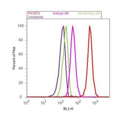
- Flow cytometry analysis of Mint3 was done on Neuro-2a cells. Cells were fixed with 70% ethanol for 10 minutes, permeabilized with 0.25% Triton™ X-100 for 20 minutes, and blocked with 5% BSA for 30 minutes at room temperature. Cells were labeled with Mint3 Rabbit Polyclonal Antibody (PA1-072, red histogram) or with rabbit isotype control (pink histogram) at 3-5 µg/million cells in 2.5% BSA. After incubation at room temperature for 2 hours, the cells were labeled with Alexa Fluor® 488 Goat Anti-Rabbit Secondary Antibody (A11008) at a dilution of 1:400 for 30 minutes at room temperature. The representative 10,000 cells were acquired and analyzed for each sample using an Attune® Acoustic Focusing Cytometer. The purple histogram represents unstained control cells and the green histogram represents no-primary-antibody control..
Supportive validation
- Submitted by
- Invitrogen Antibodies (provider)
- Main image

- Experimental details
- NULL