Explore
 Validate
 Learn
Learn Western blot
 Immunocytochemistry
Antibody data
- Antibody Data
- Antigen structure
- References [1]
- Comments [0]
- Validations
- Immunocytochemistry [3]
- Immunohistochemistry [4]
- Other assay [1]
Submit
Validation data
Reference
Comment
Report error
- Product number
- PA5-58113 - Provider product page

- Provider
- Invitrogen Antibodies
- Product name
- SNX18 Polyclonal Antibody
- Antibody type
- Polyclonal
- Antigen
- Recombinant protein fragment
- Description
- Immunogen sequence: DDEWDDSSTV ADEPGALGSG AYPDLDGSSS AGVGAAGRYR LSTRSDLSLG SRGGSVPPQH HPSGPKSSAT VSRNLN Highest antigen sequence identity to the following orthologs: Mouse - 91%, Rat - 91%.
- Reactivity
- Human, Mouse, Rat
- Host
- Rabbit
- Isotype
- IgG
- Vial size
- 100 μL
- Concentration
- 0.1 mg/mL
- Storage
- Store at 4°C short term. For long term storage, store at -20°C, avoiding freeze/thaw cycles.
Submitted references N-WASP Control of LPAR1 Trafficking Establishes Response to Self-Generated LPA Gradients to Promote Pancreatic Cancer Cell Metastasis.
Juin A, Spence HJ, Martin KJ, McGhee E, Neilson M, Cutiongco MFA, Gadegaard N, Mackay G, Fort L, Lilla S, Kalna G, Thomason P, Koh YWH, Norman JC, Insall RH, Machesky LM
Developmental cell 2019 Nov 18;51(4):431-445.e7
Developmental cell 2019 Nov 18;51(4):431-445.e7
No comments: Submit comment
Supportive validation
- Submitted by
- Invitrogen Antibodies (provider)
- Main image
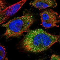
- Experimental details
- Immunofluorescent staining of SNX18 in human cell line U-251 MG shows positivity in vesicles & nucleus but excluded from the nucleoli. Samples were probed using a SNX18 Polyclonal Antibody (Product # PA5-58113).

- Submitted by
- Invitrogen Antibodies (provider)
- Main image

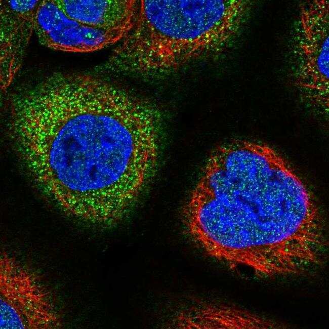
- Experimental details
- Immunofluorescent staining of SNX18 in human cell line A-431 using a SNX18 Polyclonal Antibody (Product # PA5-58113) shows localization to cytosol.
- Submitted by
- Invitrogen Antibodies (provider)
- Main image

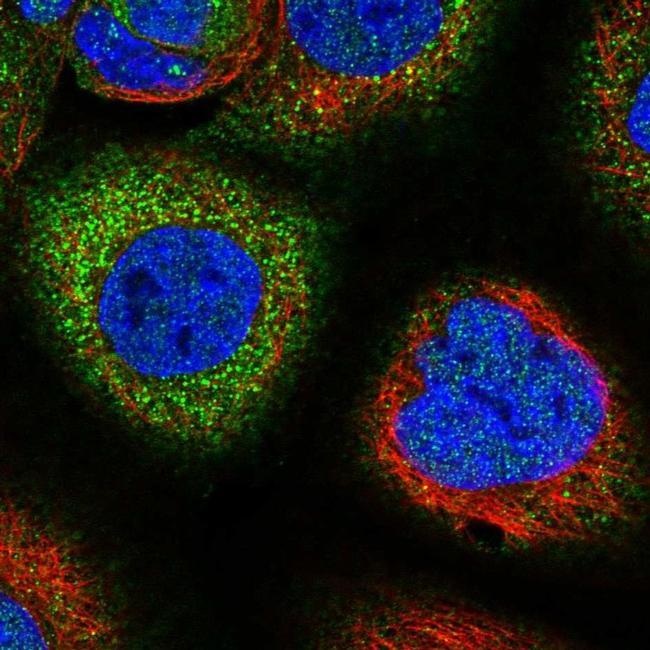
- Experimental details
- Immunofluorescent staining of SNX18 in human cell line A-431 using a SNX18 Polyclonal Antibody (Product # PA5-58113) shows localization to cytosol.
Supportive validation
- Submitted by
- Invitrogen Antibodies (provider)
- Main image
- Experimental details
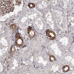
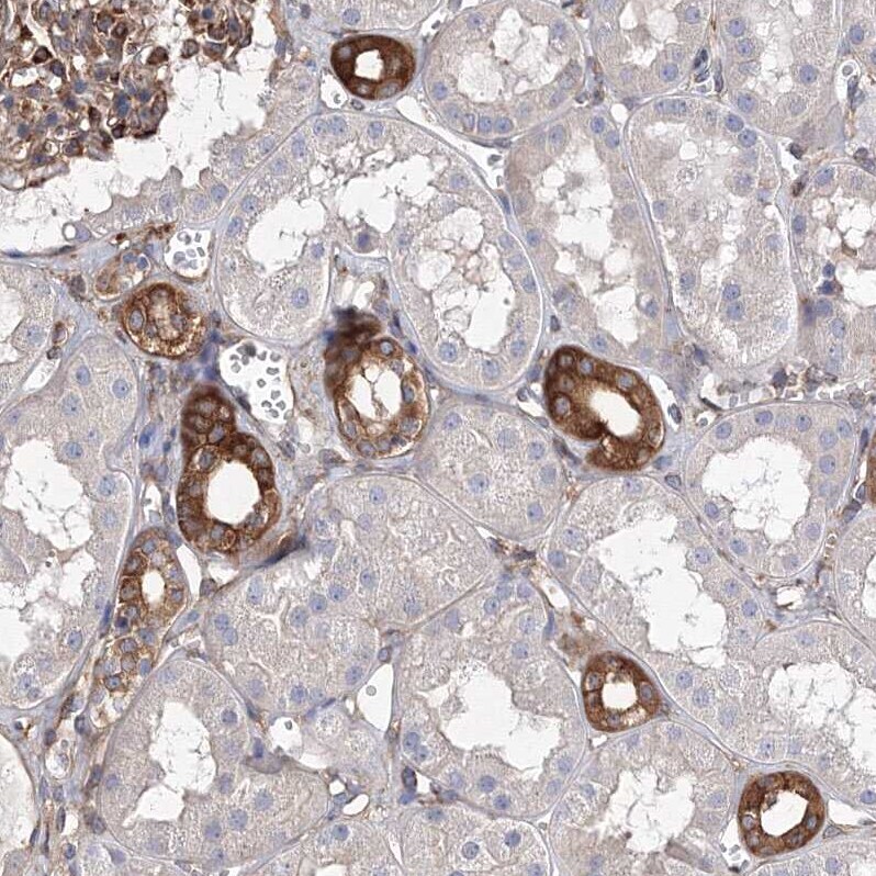
- Immunohistochemical analysis of SNX18 in human kidney using SNX18 Polyclonal Antibody (Product # PA5-58113) shows strong cytoplasmic positivity in cells in tubules.
- Submitted by
- Invitrogen Antibodies (provider)
- Main image
- Experimental details
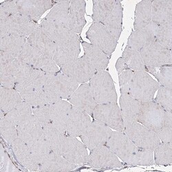
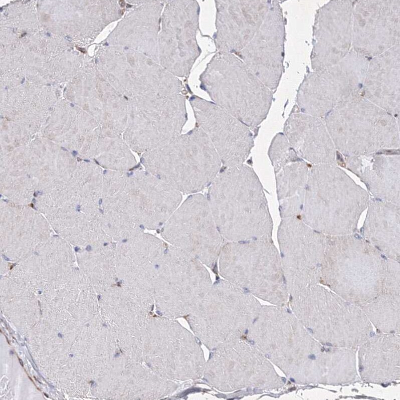
- Immunohistochemical analysis of SNX18 in human skeletal muscle using SNX18 Polyclonal Antibody (Product # PA5-58113) shows no positivity in myocytes as expected.
- Submitted by
- Invitrogen Antibodies (provider)
- Main image
- Experimental details
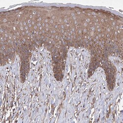
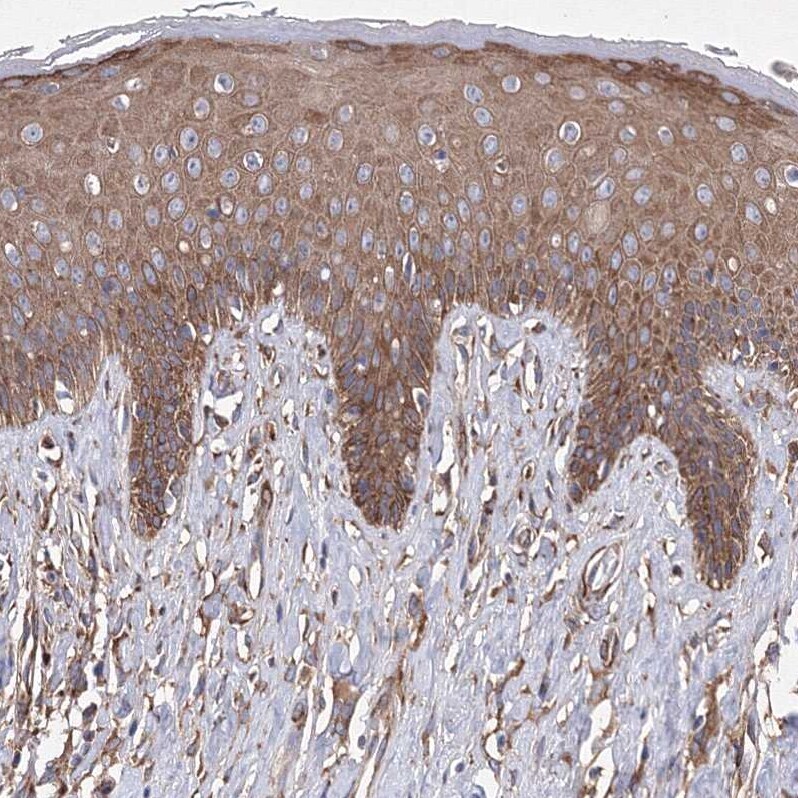
- Immunohistochemical analysis of SNX18 in human skin using SNX18 Polyclonal Antibody (Product # PA5-58113) shows strong cytoplasmic positivity in squamous epithelial cells.
- Submitted by
- Invitrogen Antibodies (provider)
- Main image
- Experimental details
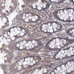
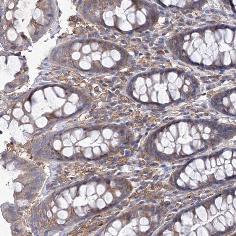
- Immunohistochemical analysis of SNX18 in human rectum using SNX18 Polyclonal Antibody (Product # PA5-58113) shows strong cytoplasmic positivity in lymphoid cells.
Supportive validation
- Submitted by
- Invitrogen Antibodies (provider)
- Main image

- Experimental details
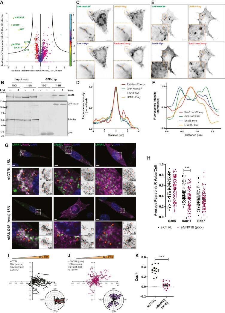
- Figure 4 The N-WASP-SNX18 Complex Regulates LPAR1 Recycling and Chemotaxis in PDAC Cells (A) Volcano plot showing proteins binding to GFP-N-WASP in 15N (rescued cells) versus GFP in 15G (N-WASP knockout cells) after 15 min LPA stimulation (t test, p