Explore
 Validate
 Learn
Learn Western blot
Antibody data
- Antibody Data
- Antigen structure
- References [3]
- Comments [0]
- Validations
- Western blot [1]
- Blocking/Neutralizing [1]
Submit
Validation data
Reference
Comment
Report error
- Product number
- AF1810 - Provider product page

- Provider
- Novus Biologicals
- Product name
- Goat Polyclonal Endocan/ESM-1 Antibody
- Antibody type
- Polyclonal
- Description
- Antigen Affinity-purified. Detects human Endocan in direct ELISAs and Western blots. In direct ELISAs and Western blots, approximately 20% cross-reactivity with recombinant mouse Endocan is observed.
- Reactivity
- Human
- Host
- Goat
- Conjugate
- Unconjugated
- Isotype
- IgG
- Vial size
- 100 ug
- Concentration
- LYOPH
- Storage
- Use a manual defrost freezer and avoid repeated freeze-thaw cycles. 12 months from date of receipt, -20 to -70 degreesC as supplied. 1 month, 2 to 8 degreesC under sterile conditions after reconstitution. 6 months, -20 to -70 degreesC under sterile conditions after reconstitution.
Submitted references Upregulation of endocan by Epstein-Barr virus latent membrane protein 1 and its clinical significance in nasopharyngeal carcinoma.
Transcriptional profiling of VEGF-A and VEGF-C target genes in lymphatic endothelium reveals endothelial-specific molecule-1 as a novel mediator of lymphangiogenesis.
Endocan is a VEGF-A and PI3K regulated gene with increased expression in human renal cancer.
Yu PH, Chou SF, Chen CL, Hung H, Lai CY, Yang PM, Jeng YM, Liaw SF, Kuo HH, Hsu HC, Chen JY, Wang WB
PloS one 2013;8(12):e82254
PloS one 2013;8(12):e82254
Transcriptional profiling of VEGF-A and VEGF-C target genes in lymphatic endothelium reveals endothelial-specific molecule-1 as a novel mediator of lymphangiogenesis.
Shin JW, Huggenberger R, Detmar M
Blood 2008 Sep 15;112(6):2318-26
Blood 2008 Sep 15;112(6):2318-26
Endocan is a VEGF-A and PI3K regulated gene with increased expression in human renal cancer.
Rennel E, Mellberg S, Dimberg A, Petersson L, Botling J, Ameur A, Westholm JO, Komorowski J, Lassalle P, Cross MJ, Gerwins P
Experimental cell research 2007 Apr 15;313(7):1285-94
Experimental cell research 2007 Apr 15;313(7):1285-94
No comments: Submit comment
Supportive validation
- Submitted by
- Novus Biologicals (provider)
- Main image
- Experimental details
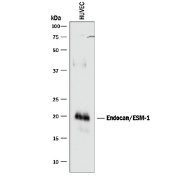
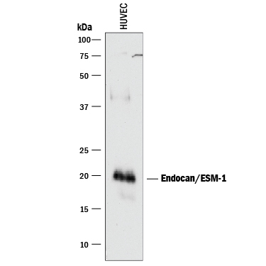
- Detection of Human Endocan/ESM-1 by Western Blot. Western blot shows lysate of HUVEC human umbilical vein endothelial cells. PVDF membrane was probed with 1 µg/mL of Goat Anti-Human Endocan/ESM-1 Antigen Affinity-purified Polyclonal Antibody (Catalog # AF1810) followed by HRP-conjugated Anti-Goat IgG Secondary Antibody (Catalog # HAF017). A specific band was detected for Endocan/ESM-1 at approximately 20 kDa (as indicated). This experiment was conducted under reducing conditions and using Immunoblot Buffer Group 1.
Supportive validation
- Submitted by
- Novus Biologicals (provider)
- Main image
- Experimental details
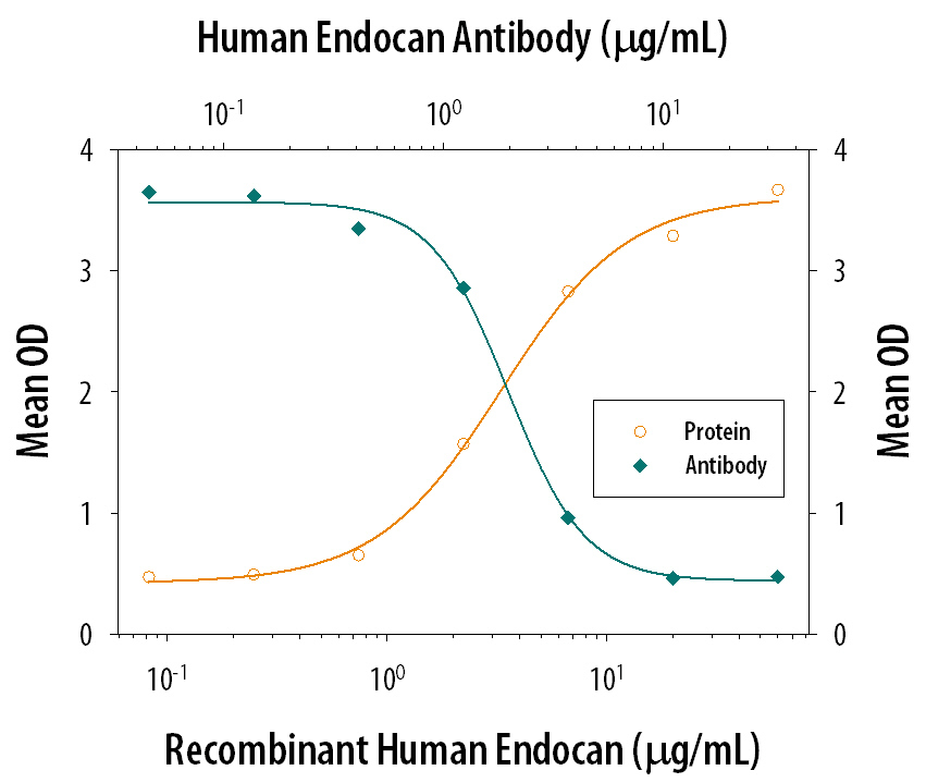
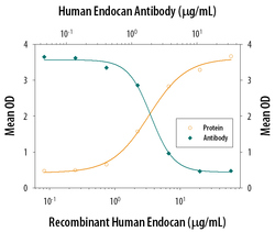
- Cell Adhesion Mediated by Endocan/ESM-1 and Neutral-ization by Human Endocan/ESM-1 Antibody. Recombinant Human Endocan/ESM-1 (Catalog # 1810-EC), immobilized onto a microplate, supports the adhesion of the Jurkat human acute T cell leukemia cell line in a dose-dependent manner (orange line). Adhesion elicited by Recombinant Human Endocan/ESM-1 (20 µg/mL) is neutralized (green line) by increasing concentrations of Goat Anti-Human Endocan/ESM-1 Antigen Affinity-purified Polyclonal Antibody (Catalog # AF1810). The ND50 is typically 1-4 µg/mL.