Explore
 Validate
 Learn
Learn Western blot
 Immunohistochemistry
Antibody data
- Antibody Data
- Antigen structure
- References [3]
- Comments [0]
- Validations
- Immunohistochemistry [1]
- Blocking/Neutralizing [1]
Submit
Validation data
Reference
Comment
Report error
- Product number
- AF1999 - Provider product page

- Provider
- R&D Systems
- Product name
- Mouse Endocan/ESM-1 Antibody
- Antibody type
- Polyclonal
- Description
- Antigen Affinity-purified. Detects mouse Endocan/ESM-1 in direct ELISAs and Western blots. In direct ELISAs and Western blots, approximately 25% cross-reactivity with recombinant human Endocan is observed.
- Reactivity
- Mouse
- Host
- Goat
- Conjugate
- Unconjugated
- Antigen sequence
Q9QYY7- Isotype
- IgG
- Vial size
- 100 ug
- Concentration
- LYOPH
- Storage
- Use a manual defrost freezer and avoid repeated freeze-thaw cycles. 12 months from date of receipt, -20 to -70 °C as supplied. 1 month, 2 to 8 °C under sterile conditions after reconstitution. 6 months, -20 to -70 °C under sterile conditions after reconstitution.
Submitted references Endocan Blockade Suppresses Experimental Ocular Neovascularization in Mice.
Integrin β1 controls VE-cadherin localization and blood vessel stability.
Transcriptional profiling of VEGF-A and VEGF-C target genes in lymphatic endothelium reveals endothelial-specific molecule-1 as a novel mediator of lymphangiogenesis.
Su T, Zhong Y, Demetriades AM, Shen J, Sui A, Yao Y, Gao Y, Zhu Y, Shen X, Xie B
Investigative ophthalmology & visual science 2018 Feb 1;59(2):930-939
Investigative ophthalmology & visual science 2018 Feb 1;59(2):930-939
Integrin β1 controls VE-cadherin localization and blood vessel stability.
Yamamoto H, Ehling M, Kato K, Kanai K, van Lessen M, Frye M, Zeuschner D, Nakayama M, Vestweber D, Adams RH
Nature communications 2015 Mar 10;6:6429
Nature communications 2015 Mar 10;6:6429
Transcriptional profiling of VEGF-A and VEGF-C target genes in lymphatic endothelium reveals endothelial-specific molecule-1 as a novel mediator of lymphangiogenesis.
Shin JW, Huggenberger R, Detmar M
Blood 2008 Sep 15;112(6):2318-26
Blood 2008 Sep 15;112(6):2318-26
No comments: Submit comment
Supportive validation
- Submitted by
- R&D Systems (provider)
- Main image

- Experimental details
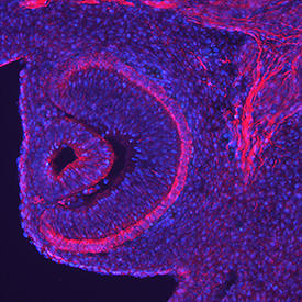
- Endocan/ESM-1 in Mouse Embryo. Endocan/ESM-1 was detected in perfusion fixed frozen sections of mouse embryo (13 d.p.c.) using Goat Anti-Mouse Endocan/ESM-1 Antigen Affinity-purified Polyclonal Antibody (Catalog # AF1999) at 5 µg/mL overnight at 4 °C. Tissue was stained using the NorthernLights™ 557-conjugated Anti-Goat IgG Secondary Antibody (red; Catalog # NL001) and counterstained with DAPI (blue). Specific staining was localized to the retina. View our protocol for Fluorescent IHC Staining of Frozen Tissue Sections.
Supportive validation
- Submitted by
- R&D Systems (provider)
- Main image
- Experimental details
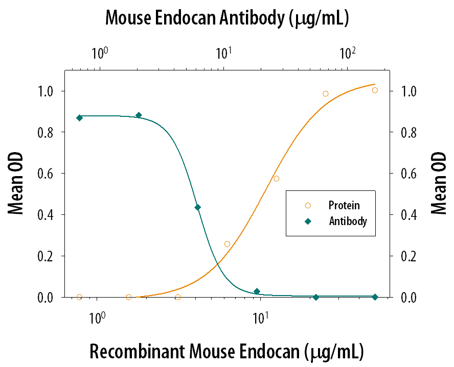
- Cell Adhesion Mediated by Endocan/ESM-1 and Neutralization by Mouse Endocan/ESM-1 Antibody. Recombinant Mouse Endocan/ESM-1 (Catalog # 1999-EC), immobilized onto a microplate, supports the adhesion of the Jurkat human acute T cell leukemia cell line in a dose-dependent manner (orange line). Adhesion elicited by Recombinant Mouse Endocan/ESM-1 (25 µg/mL) is neutralized (green line) by increasing concentrations of Goat Anti-Mouse Endocan/ESM-1 Antigen Affinity-purified Polyclonal Antibody (Catalog # AF1999). The ND50 is typically 1-5 µg/mL.