Explore
 Validate
 Learn
Learn Western blot
 Immunocytochemistry
Antibody data
- Antibody Data
- Antigen structure
- References [4]
- Comments [0]
- Validations
- Immunocytochemistry [2]
- Other assay [5]
Submit
Validation data
Reference
Comment
Report error
- Product number
- PA1-732 - Provider product page

- Provider
- Invitrogen Antibodies
- Product name
- beta-Arrestin 2 Polyclonal Antibody
- Antibody type
- Polyclonal
- Antigen
- Synthetic peptide
- Description
- PA1-732 detects beta-Arrestin 2 in rat, human and bovine samples. PA1-732 has been successfully used in Western blot procedures. By Western blot, this antibody detects ~49 kDa and ~47 kDa proteins representing pan Arrestin in rat and bovine retina as well as rat brain samples. The PA1-732 immunogen is a synthetic peptide corresponding to residues C D(384) D I V F E D F A R L R L K(397) of human beta-arrestin 2. This peptide (Cat. # PEP-281) is available for use in neutralization and control experiments.
- Reactivity
- Human, Mouse, Rat, Bovine
- Host
- Rabbit
- Isotype
- IgG
- Vial size
- 100 μg
- Concentration
- 1 mg/mL
- Storage
- -20°C, Avoid Freeze/Thaw Cycles
Submitted references Colocalization and Interaction Study of Neuronal JNK3, JIP1, and β-Arrestin2 Together with PSD95.
Alterations of the renin angiotensin system in human end-stage heart failure before and after mechanical cardiac unloading by LVAD support.
Arrestin competition influences the kinetics and variability of the single-photon responses of mammalian rod photoreceptors.
Dopamine promotes striatal neuronal apoptotic death via ERK signaling cascades.
Musi CA, Marchini G, Giani A, Tomaselli G, Priori EC, Colnaghi L, Borsello T
International journal of molecular sciences 2022 Apr 8;23(8)
International journal of molecular sciences 2022 Apr 8;23(8)
Alterations of the renin angiotensin system in human end-stage heart failure before and after mechanical cardiac unloading by LVAD support.
Messmann R, Dietl A, Wagner S, Domenig O, Jungbauer C, Luchner A, Maier LS, Schopka S, Hirt S, Schmid C, Birner C
Molecular and cellular biochemistry 2020 Sep;472(1-2):79-94
Molecular and cellular biochemistry 2020 Sep;472(1-2):79-94
Arrestin competition influences the kinetics and variability of the single-photon responses of mammalian rod photoreceptors.
Doan T, Azevedo AW, Hurley JB, Rieke F
The Journal of neuroscience : the official journal of the Society for Neuroscience 2009 Sep 23;29(38):11867-79
The Journal of neuroscience : the official journal of the Society for Neuroscience 2009 Sep 23;29(38):11867-79
Dopamine promotes striatal neuronal apoptotic death via ERK signaling cascades.
Chen J, Rusnak M, Lombroso PJ, Sidhu A
The European journal of neuroscience 2009 Jan;29(2):287-306
The European journal of neuroscience 2009 Jan;29(2):287-306
No comments: Submit comment
Supportive validation
- Submitted by
- Invitrogen Antibodies (provider)
- Main image

- Experimental details
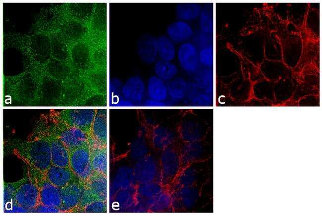
- Immunofluorescence analysis of beta-Arrestin 2 was performed using 70% confluent log phase HEK293 cells. The cells were fixed with 4% paraformaldehyde for 10 minutes, permeabilized with 0.1% Triton™ X-100 for 10 minutes, and blocked with 1% BSA for 1 hour at room temperature. The cells were labeled with beta-Arrestin 2 Rabbit Polyclonal Antibody (Product # PA1-732) at 2µg/mL in 0.1% BSA and incubated for 3 hours at room temperature and then labeled with Goat anti-Rabbit IgG (H+L) Superclonal™ Secondary Antibody, Alexa Fluor® 488 conjugate (Product # A27034) at a dilution of 1:2000 for 45 minutes at room temperature (Panel a: green). Nuclei (Panel b: blue) were stained with SlowFade® Gold Antifade Mountant with DAPI (Product # S36938). F-actin (Panel c: red) was stained with Rhodamine Phalloidin (Product # R415, 1:300). Panel d represents the merged image showing cytoplasmic localization. Panel e shows the no primary antibody control. The images were captured at 60X magnification.
- Submitted by
- Invitrogen Antibodies (provider)
- Main image

- Experimental details
- Immunofluorescence analysis of beta-Arrestin 2 was performed using 70% confluent log phase HEK293 cells. The cells were fixed with 4% paraformaldehyde for 10 minutes, permeabilized with 0.1% Triton™ X-100 for 10 minutes, and blocked with 1% BSA for 1 hour at room temperature. The cells were labeled with beta-Arrestin 2 Rabbit Polyclonal Antibody (Product # PA1-732) at 2µg/mL in 0.1% BSA and incubated for 3 hours at room temperature and then labeled with Goat anti-Rabbit IgG (Heavy Chain) Superclonal™ Secondary Antibody, Alexa Fluor® 488 conjugate (Product # A27034) at a dilution of 1:2000 for 45 minutes at room temperature (Panel a: green). Nuclei (Panel b: blue) were stained with SlowFade® Gold Antifade Mountant with DAPI (Product # S36938). F-actin (Panel c: red) was stained with Rhodamine Phalloidin (Product # R415, 1:300). Panel d represents the merged image showing cytoplasmic localization. Panel e shows the no primary antibody control. The images were captured at 60X magnification.

Supportive validation
- Submitted by
- Invitrogen Antibodies (provider)
- Main image
- Experimental details
- NULL

- Submitted by
- Invitrogen Antibodies (provider)
- Main image

- Experimental details
- NULL
- Submitted by
- Invitrogen Antibodies (provider)
- Main image

- Experimental details
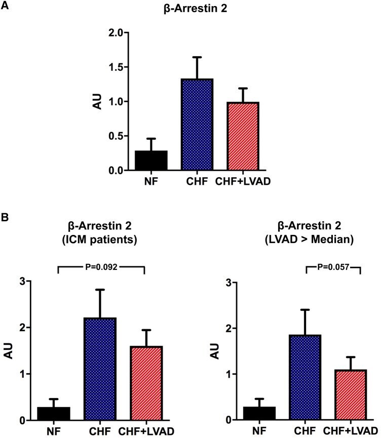
- Fig. 8 a beta-arrestin 2 before (CHF) and after LVAD therapy (CHF+LVAD) as compared to non-failing ventricles (NF). beta-arrestin 2 expression was determined by immunoblot (western blot) analysis and referred to a standard, respectively, whose densitometric value was set 1 by default. The value on the y -axis, therefore, reflects the percentage of each parameter's immunoblot band density in relation to this default value. AU arbitrary unit, CHF congestive heart failure, LVAD left ventricular assist device, NF non-failing myocardial tissue specimen. b beta-arrestin 2 before (CHF) and after LVAD therapy (CHF+LVAD) in the subgroup of ICM patients as compared to non-failing ventricles (NF), left ( n = 8). beta-arrestin 2 before (CHF) and after LVAD therapy (CHF+LVAD) in the subgroup of 10 patients with a duration of LVAD therapy above the median value as compared to non-failing ventricles (NF), right. AU arbitrary unit, CHF congestive heart failure, ICM ischemic cardiomyopathy, LVAD left ventricular assist device, NF non-failing myocardial tissue specimen
- Submitted by
- Invitrogen Antibodies (provider)
- Main image
- Experimental details
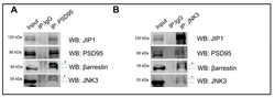
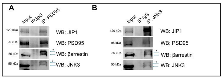
- Immunoprecipitation outputs used to assess JNK3-JIP1-beta-arrestin2-PSD95 colocalization in primary hippocampal neurons. ( A ) PSD95 was immunoprecipitated from the cell lysate homogenate using PSD95 antibody, and immune complexes were analyzed for the presence of JNK3, JIP1, and beta-arrestin2. Immunoprecipitation with IgG antibody was used as a control. ( B ) JNK3 was immunoprecipitated from the cell lysate homogenate using JNK3-specific antibody, separated by SDS-PAGE, and analyzed via Western blot with anti-PSD95, anti-JIP1, and anti-beta-arrestin2 antibodies. Immunoprecipitation with IgG antibody was used as a control. * indicates non-specific bands.
- Submitted by
- Invitrogen Antibodies (provider)
- Main image
- Experimental details
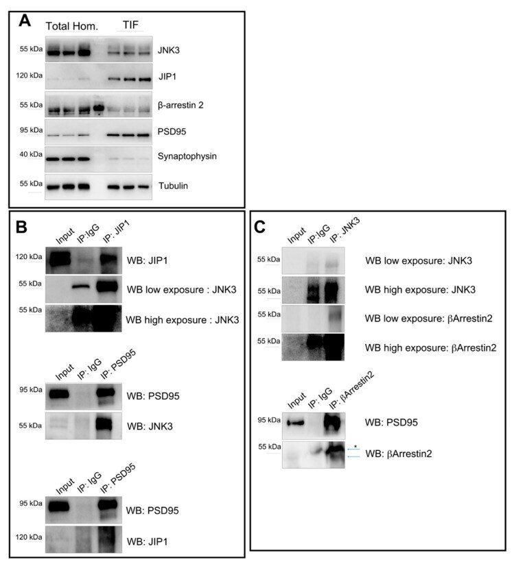
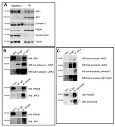
- Immunoprecipitation outputs used to assess JNK3-JIP1-beta-arrestin2-PSD95 interactions at the synapse. ( A ) Western blots performed on total brain homogenate and Triton-insoluble fraction (TIF) reveal the different amounts of JNK3, JIP1, beta-arrestin2, and PSD95 in the two extracts analyzed. Synaptophysin was used as a control for the purity of the TIF preparation. ( B ) JIP1 was immunoprecipitated from the TIF using JIP1 antibody, and immune complexes were analyzed for the presence of JNK3 (rabbit). Immunoprecipitation with IgG antibody was used as a control. PSD95 was immunoprecipitated from the TIF using PSD95-specific antibody, separated by SDS-PAGE, and analyzed with Western blot with anti-PSD95, anti-JNK3 (mouse), and anti-JIP1 antibodies. Immunoprecipitation with IgG antibody was used as a control. ( C ) JNK3 was immunoprecipitated from the TIF using the JNK3 antibody, and immune complexes were analyzed for the presence of beta-arrestin2 and JNK3 (rabbit). Immunoprecipitation with IgG antibody was used as a control. beta-arrestin2 was immunoprecipitated from the TIF using beta-arrestin2-specific antibody, separated by SDS-PAGE, and analyzed by Western blot with anti-PSD95, anti-JNK3, and anti-beta-arrestin2 antibodies. Immunoprecipitation with IgG antibody was used as a control. * indicates non-specific bands.