Explore
 Validate
 Learn
Learn Western blot
Antibody data
- Antibody Data
- Antigen structure
- References [0]
- Comments [0]
- Validations
- Western blot [4]
- Immunohistochemistry [1]
Submit
Validation data
Reference
Comment
Report error
- Product number
- PA5-23296 - Provider product page

- Provider
- Invitrogen Antibodies
- Product name
- beta-Arrestin 2 Polyclonal Antibody
- Antibody type
- Polyclonal
- Antigen
- Synthetic peptide
- Reactivity
- Human, Mouse, Rat, Bovine, Canine, Chicken/Avian, Xenopus, Zebrafish
- Host
- Rabbit
- Isotype
- IgG
- Vial size
- 100 µg
- Concentration
- 0.5 mg/mL
- Storage
- -20° C, Avoid Freeze/Thaw Cycles
No comments: Submit comment
Supportive validation
- Submitted by
- Invitrogen Antibodies (provider)
- Main image
- Experimental details
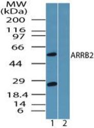
- Western blot analysis of ARRB2 in HUVEC cell lysate in the 1) absence and 2) presence of immunizing peptide using a ARRB2 polyclonal antibody (Product # PA5-23296) at 0.25 µg/mL. Goat anti-rabbit Ig HRP secondary antibody.

- Submitted by
- Invitrogen Antibodies (provider)
- Main image

- Experimental details
- Western blot analysis of beta-Arrestin 2 in HUVEC cell lysate in the 1) absence and 2) presence of immunizing peptide. Samples were incubated in beta-Arrestin 2 polyclonal antibody (Product # PA5-23296) using a dilution of 0.25 µg/mL followed by a goat anti-rabbit Ig HRP secondary antibody. PicoTect ECL substrate solution was used for this test.

- Submitted by
- Invitrogen Antibodies (provider)
- Main image

- Experimental details
- Western blot analysis of beta-Arrestin 2 in 0.5 mg/mL Jurkat lysate. Samples were incubated in beta-Arrestin 2 polyclonal antibody (Product # PA5-23296). This experiment was performed under reducing conditions using the 12-230 kDa separation system.

- Submitted by
- Invitrogen Antibodies (provider)
- Main image
- Experimental details
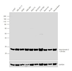
- Western blot was performed using Anti-beta-Arrestin 2 Polyclonal Antibody (Product # PA5-23296) and a 42 kDa band corresponding to beta-Arrestin 2 was observed across cell lines tested. Whole cell extracts (30 µg lysate) of U-937 (Lane 1), HEL 92.1.7 (Lane 2), A549 (Lane 3), HEK-293 (Lane 4), MOLT-4 (Lane 5), SH-SY5Y (Lane 6), U-87 MG (Lane 7), Neuro-2a (Lane 8), HL-60 (Lane 9) and tissue extract (30 µg lysate) of Mouse Brain (Lane 10) were electrophoresed using NuPAGE™ 10% Bis-Tris Protein Gel (Product # NP0302BOX). Resolved proteins were then transferred onto a nitrocellulose membrane (Product # IB23001) by iBlot® 2 Dry Blotting System (Product # IB21001). The blot was probed with the primary antibody (1 µg/mL) and detected by chemiluminescence Goat Anti-Rabbit IgG Secondary Antibody, HRP conjugate (Product # A27036, 1:4000 dilution) using the iBright FL 1000 (Product # A32752). Chemiluminescent detection was performed using Novex® ECL Chemiluminescent Substrate Reagent Kit (Product # WP20005)..
Supportive validation
- Submitted by
- Invitrogen Antibodies (provider)
- Main image

- Experimental details
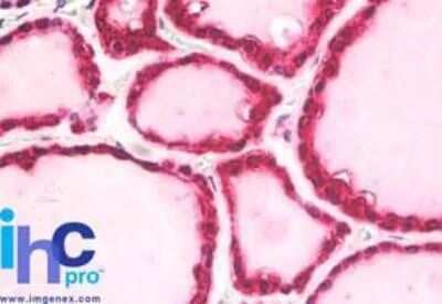
- Immunohistochemical analysis of beta-Arrestin 2 in Human thyroid tissue. Samples were incubated with beta-Arrestin 2 polyclonal antibody (Product # PA5-23296) using a dilution of 5 µg/mL. Staining of formalin-fixed tissues is enhanced by boiling tissue sections in 10 mM sodium citrate buffer, pH 6.0 for 10-20 min followed by cooling at RT for 20 min.