 Explore
 Validate
 Learn
LearnAP09300PU-N
antibody from Acris Antibodies GmbH
Targeting: HMGN1
FLJ27265, FLJ31471, HMG14, MGC104230, MGC117425
 Western blot
 ELISA
ELISAAntibody data
- Antibody Data
- Antigen structure
- References [0]
- Comments [0]
- Validations
- Western blot [1]
- Immunohistochemistry [1]
Submit
Validation data
Reference
Comment
Report error
- Product number
- AP09300PU-N - Provider product page

- Provider
- Acris Antibodies GmbH
- Proper citation
- Acris Antibodies GmbH Cat#AP09300PU-N, RRID:AB_2035640
- Product name
- anti HMGN1 / HMGN2 pSer20/24
- Antibody type
- Polyclonal
- Antigen
- Synthetic peptide corresponding to amino acids 19-28 of human HMGN protein
- Reactivity
- Human, Mouse, Rat, Bovine, Canine, Chicken/Avian, Porcine, Xenopus
- Host
- Rabbit
- Isotype
- IgG
- Vial size
- 0.1 mg
- Concentration
- 1.20 mg/ml (by UV absorbance at 280 nm)
No comments: Submit comment
Supportive validation
- Submitted by
- Acris Antibodies GmbH (provider)
- Main image

- Experimental details
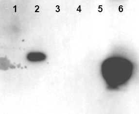
- Western blot using Affinity Purified anti-HMNG antibody shows detection of phosphorylated HMGN1 and HMGN2. Recombinant native and mutant HMGN proteins were treated with kinase PKCalpha to specifically phosphorylate S20 and S24 residues. Lanes contain: 1 - HMGN1, nonphosphorylated, 2 - HMGN1, phosphorylated, 3 - HMGN1 delta20E, delta24E, non- phosphorylated, 4 - HMGN1, delta20E, delta24E, phosphorylated, 5 - HMGN2, non-phosphorylated, and 6 - HMGN2, phosphorylated. Molecular weight markers (not shown) confirm the size of each recombinant protein. The primary antibody was diluted 1:1,000 for this experiment. The blot was processed using a 5 sec exposure time.
Supportive validation
- Submitted by
- Acris Antibodies GmbH (provider)
- Main image
- Experimental details
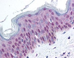
- Immunohistochemistry. affinity purified anti-HMGN pS20/pS24 antibody was used at 20 µg/ml to detectsignal in a variety of tissues including multi-human, multi-brain and multi-cancer slides. This image shows moderate nuclear and faint cytoplasmic positive staining of epidermal keratinocytes at 40X. Tissue was formalin-fixed and paraffin embedded. The image shows localization of the antibody as the precipitated red signal, with a hematoxylin purple nuclear counterstain.
