 Explore
 Validate
 Learn
Learn Western blot
Antibody data
- Antibody Data
- Antigen structure
- References [2]
- Comments [0]
- Validations
- Western blot [1]
- Immunocytochemistry [3]
- Immunoprecipitation [1]
- Immunohistochemistry [8]
- Other assay [2]
Submit
Validation data
Reference
Comment
Report error
- Product number
- PA5-29199 - Provider product page

- Provider
- Invitrogen Antibodies
- Product name
- SLC25A6 Polyclonal Antibody
- Antibody type
- Polyclonal
- Antigen
- Recombinant full-length protein
- Description
- Recommended positive controls: 293T. Predicted reactivity: Zebrafish (93%), Pig (96%), Chicken (93%), Sheep (97%), Bovine (97%). Store product as a concentrated solution. Centrifuge briefly prior to opening the vial.
- Reactivity
- Human, Mouse, Rat
- Host
- Rabbit
- Isotype
- IgG
- Vial size
- 100 μL
- Concentration
- 1.7 mg/mL
- Storage
- Store at 4°C short term. For long term storage, store at -20°C, avoiding freeze/thaw cycles.
Submitted references The RNA-Binding Protein YBX3 Controls Amino Acid Levels by Regulating SLC mRNA Abundance.
New interaction partners for Nek4.1 and Nek4.2 isoforms: from the DNA damage response to RNA splicing.
Cooke A, Schwarzl T, Huppertz I, Kramer G, Mantas P, Alleaume AM, Huber W, Krijgsveld J, Hentze MW
Cell reports 2019 Jun 11;27(11):3097-3106.e5
Cell reports 2019 Jun 11;27(11):3097-3106.e5
New interaction partners for Nek4.1 and Nek4.2 isoforms: from the DNA damage response to RNA splicing.
Basei FL, Meirelles GV, Righetto GL, Dos Santos Migueleti DL, Smetana JH, Kobarg J
Proteome science 2015;13:11
Proteome science 2015;13:11
No comments: Submit comment
Supportive validation
- Submitted by
- Invitrogen Antibodies (provider)
- Main image
- Experimental details
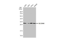
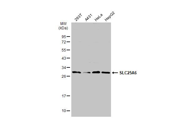
- Western Blot using SLC25A6 Polyclonal Antibody (Product # PA5-29199). Various whole cell extracts (30 µg) were separated by 12% SDS-PAGE, and the membrane was blotted with SLC25A6 Polyclonal Antibody (Product # PA5-29199) diluted at 1:2,000. The HRP-conjugated anti-rabbit IgG antibody was used to detect the primary antibody.
Supportive validation
- Submitted by
- Invitrogen Antibodies (provider)
- Main image
- Experimental details
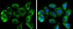
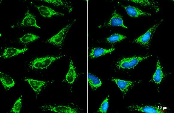
- Immunocytochemistry-Immunofluorescence analysis of SLC25A6 was performed in MCF7 cells fixed in 4% paraformaldehyde at RT for 15 min. Green: SLC25A6 Polyclonal Antibody (Product # PA5-29199) diluted at 1:500. Blue: Hoechst 33342 staining.

- Submitted by
- Invitrogen Antibodies (provider)
- Main image
- Experimental details
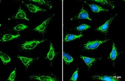
- SLC25A6 Polyclonal Antibody detects SLC25A6 protein at mitochondria by immunofluorescent analysis. Sample: HeLa cells were fixed in ice-cold MeOH for 5 min. Green: SLC25A6 stained by SLC25A6 Polyclonal Antibody (Product # PA5-29199) diluted at 1:500. Blue: Fluoroshield with DAPI . Scale bar= 10 µm.

- Submitted by
- Invitrogen Antibodies (provider)
- Main image

- Experimental details
- SLC25A6 Polyclonal Antibody detects SLC25A6 protein at mitochondria by immunofluorescent analysis. Sample: HeLa cells were fixed in ice-cold MeOH for 5 min. Green: SLC25A6 stained by SLC25A6 Polyclonal Antibody (Product # PA5-29199) diluted at 1:500. Blue: Fluoroshield with DAPI . Scale bar= 10 µm.
Supportive validation
- Submitted by
- Invitrogen Antibodies (provider)
- Main image

- Experimental details
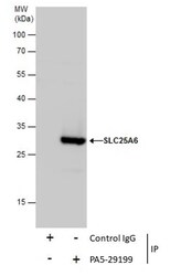
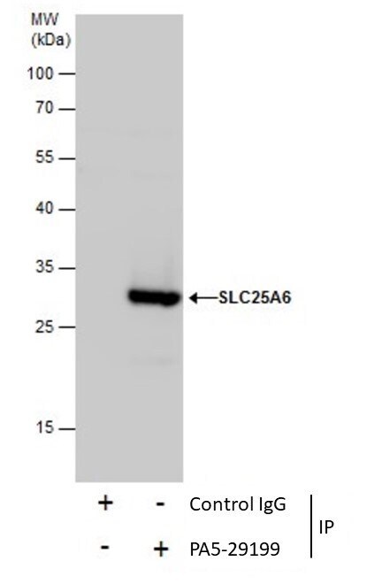
- Immunoprecipitation of SLC25A6 was performed in 293T whole cell extracts using 5 µg of SLC25A6 Polyclonal Antibody (Product # PA5-29199). Samples were transferred to a membrane and probed with SLC25A6 Polyclonal Antibody as a primary antibody and an HRP-conjugated anti-Rabbit IgG was used as a secondary antibody.

Supportive validation
- Submitted by
- Invitrogen Antibodies (provider)
- Main image
- Experimental details
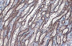
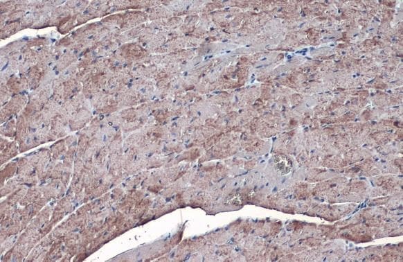
- SLC25A6 Polyclonal Antibody detects SLC25A6 protein at mitochondria by immunohistochemical analysis. Sample: Paraffin-embedded rat kidney. SLC25A6 stained by SLC25A6 Polyclonal Antibody (Product # PA5-29199) diluted at 1:1,000. Antigen Retrieval: Citrate buffer, pH 6.0, 15 min.

- Submitted by
- Invitrogen Antibodies (provider)
- Main image
- Experimental details
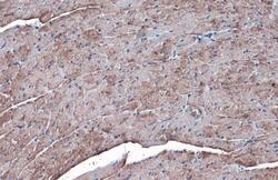
- SLC25A6 Polyclonal Antibody detects SLC25A6 protein at mitochondria by immunohistochemical analysis. Sample: Paraffin-embedded mouse heart. SLC25A6 stained by SLC25A6 Polyclonal Antibody (Product # PA5-29199) diluted at 1:1,000. Antigen Retrieval: Citrate buffer, pH 6.0, 15 min.

- Submitted by
- Invitrogen Antibodies (provider)
- Main image

- Experimental details
- SLC25A6 Polyclonal Antibody detects SLC25A6 protein at mitochondria by immunohistochemical analysis. Sample: Paraffin-embedded rat muscle. SLC25A6 stained by SLC25A6 Polyclonal Antibody (Product # PA5-29199) diluted at 1:1,000. Antigen Retrieval: Citrate buffer, pH 6.0, 15 min.

- Submitted by
- Invitrogen Antibodies (provider)
- Main image

- Experimental details
- SLC25A6 Polyclonal Antibody detects SLC25A6 protein at mitochondria by immunohistochemical analysis. Sample: Paraffin-embedded mouse heart. SLC25A6 stained by SLC25A6 Polyclonal Antibody (Product # PA5-29199) diluted at 1:1,000. Antigen Retrieval: Citrate buffer, pH 6.0, 15 min.

- Submitted by
- Invitrogen Antibodies (provider)
- Main image
- Experimental details
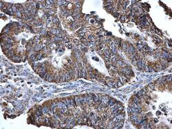
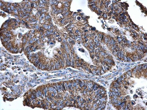
- Immunohistochemistry (Paraffin) analysis of SLC25A6 was performed in paraffin-embedded human colon cancer tissue using SLC25A6 Polyclonal Antibody (Product # PA5-29199) at a dilution of 1:500.
- Submitted by
- Invitrogen Antibodies (provider)
- Main image

- Experimental details
- SLC25A6 Polyclonal Antibody detects SLC25A6 protein at mitochondria by immunohistochemical analysis. Sample: Paraffin-embedded mouse heart. SLC25A6 stained by SLC25A6 Polyclonal Antibody (Product # PA5-29199) diluted at 1:1,000. Antigen Retrieval: Citrate buffer, pH 6.0, 15 min.

- Submitted by
- Invitrogen Antibodies (provider)
- Main image

- Experimental details
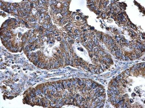
- Immunohistochemistry (Paraffin) analysis of SLC25A6 was performed in paraffin-embedded human colon cancer tissue using SLC25A6 Polyclonal Antibody (Product # PA5-29199) at a dilution of 1:500.
Supportive validation
- Submitted by
- Invitrogen Antibodies (provider)
- Main image
- Experimental details
- Immunoprecipitation of SLC25A6 was performed in 293T whole cell extracts using 5 µg of SLC25A6 Polyclonal Antibody (Product # PA5-29199). Samples were transferred to a membrane and probed with SLC25A6 Polyclonal Antibody as a primary antibody and an HRP-conjugated anti-Rabbit IgG was used as a secondary antibody.
- Submitted by
- Invitrogen Antibodies (provider)
- Main image
- Experimental details
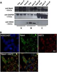
- Figure 4 Nek4 copurifies with the mitochondrial fraction. (A) Nek4 is present in mitochondrial fractions and shows a similar perinuclear pattern of localization (B) such as its putative interactor ANT3, a mitochondrial protein. HEK293 cells were seeded on Cell Carrier 384 well plates coated with polylysine (Perkin Elmer), incubated for two days at 37degC with 5% CO2, then fixed with methanol and stained with proper antibodies. The nuclei were stained with Hoechst. Images were acquired with the Operetta automated microscope (Perkin Elmer) in non-confocal mode using the 60x high NA objective (NA = 0.9). The datasets were imported into Volocity for colocalization analysis and contrast correction. The images are representative of at least 10 replicate experiments. The mitochondrial localization of Nek4 was verified by cell fractioning of HEK293 Flp-In (stably expressing Nek4 isoforms or empty vector - Flag). Using Qproteome Mitochondria Isolation Kit (QIAGEN). N: nuclear, M: mitochondrial and C: cytosolic fractions.
