 Explore
 Validate
 Learn
Learn Western blot
 ELISA
ELISAAntibody data
- Antibody Data
- Antigen structure
- References [0]
- Comments [0]
- Validations
- Western blot [4]
- Immunocytochemistry [2]
- Immunohistochemistry [2]
Submit
Validation data
Reference
Comment
Report error
- Product number
- MA5-15833 - Provider product page

- Provider
- Invitrogen Antibodies
- Product name
- EEF2 Monoclonal Antibody (5B6)
- Antibody type
- Monoclonal
- Antigen
- Purifed from natural sources
- Description
- MA5-15833 targets EEF2 in indirect ELISA, IF, IHC, and WB applications and shows reactivity with Human samples. The MA5-15833 immunogen is purified recombinant fragment of human EEF2 expressed in E. Coli. . MA5-15833 detects EEF2 which has a predicted molecular weight of approximately 95kDa.
- Reactivity
- Human, Mouse
- Host
- Mouse
- Isotype
- IgG
- Antibody clone number
- 5B6
- Vial size
- 100 µL
- Concentration
- Conc. Not Determined
- Storage
- Store at 4°C short term. For long term storage, store at -20°C, avoiding freeze/thaw cycles.
No comments: Submit comment
Supportive validation
- Submitted by
- Invitrogen Antibodies (provider)
- Main image
- Experimental details
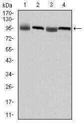
- Western blot analysis of EEF2 using EEF2 monoclonal antibody (Product # MA5-15833) in HepG2 (1), HeLa (2), HEK293 (3) and A431 (4) cell lysate.

- Submitted by
- Invitrogen Antibodies (provider)
- Main image

- Experimental details
- Western blot analysis of EEF2 using EEF2 monoclonal antibody (Product # MA5-15833) in HepG2 (1), HeLa (2), HEK293 (3) and A431 (4) cell lysate.

- Submitted by
- Invitrogen Antibodies (provider)
- Main image

- Experimental details
- Knockdown of EEF2 was achieved by transfecting HeLa cells with EEF2 specific siRNAs (Silencer® select Product # s4492). Western blot analysis (Fig. a) was performed using whole cell extracts from the EEF2 knockdown cells (lane 3), non-specific scrambled siRNA transfected cells (lane 2) and untransfected cells (lane 1). The blots were probed with EEF2 Monoclonal Antibody (5B6) (Product # MA5-15833, 1:1000 dilution) and Goat anti-Mouse IgG (H+L) Superclonal™ Secondary Antibody, HRP conjugate (Product # A28177, 0.25 µg/mL, 1:4000 dilution). Densitometric analysis of this western blot is shown in histogram (Fig. b). Decrease in signal upon siRNA mediated knock down confirms that antibody is specific to EEF2.

- Submitted by
- Invitrogen Antibodies (provider)
- Main image

- Experimental details
- Western blot analysis was performed on modified whole cell extracts (1% SDS) (30 µg lysate) of HCT 116 (Lane 1), HeLa (Lane 2), Hep G2 (Lane 3), HEK-293 (Lane 4), A-431 (Lane 5), HT-29 (Lane 6), SH-SY5Y (Lane 7), MCF7 (Lane 8), 3T3-L1 (Lane 9), A549 (Lane 10) and T-47D (Lane 11). The blot was probed with Anti-EEF2 Monoclonal Antibody (Product # MA5-15833, 1:1000 dilution) and detected by chemiluminescence using Goat anti-Mouse IgG (H+L) Superclonal™ Secondary Antibody, HRP conjugate (Product # A28177, 0.25 µg/mL, 1:4000 dilution). A 95 kDa band corresponding to EEF2 was detected across all the cell lines tested.

Supportive validation
- Submitted by
- Invitrogen Antibodies (provider)
- Main image

- Experimental details
- Immunofluorescence analysis of 3T3-L1 cells using EEF2 monoclonal antibody (Product # MA5-15833) (Green). Blue: DRAQ5 fluorescent DNA dye. Red: actin filaments have been labeled with phalloidin.

- Submitted by
- Invitrogen Antibodies (provider)
- Main image

- Experimental details
- Immunofluorescence analysis of EEF2 was performed using 70% confluent log phase HCT 116 cells. The cells were fixed with 4% paraformaldehyde for 10 minutes, permeabilized with 0.1% Triton™ X-100 for 15 minutes, and blocked with 1% BSA for 1 hour at room temperature. The cells were labeled with EEF2 Monoclonal Antibody (5B6) (Product # MA5-15833) at 1:100 dilution in 0.1% BSA, incubated at 4 degree Celsius overnight and then labeled with Goat anti-Mouse IgG (H+L) Superclonal™ Secondary Antibody, Alexa Fluor® 488 conjugate (Product # A28175) at a dilution of 1:2000 for 45 minutes at room temperature (Panel a: green). Nuclei (Panel b: blue) were stained with ProLong™ Diamond Antifade Mountant with DAPI (Product # P36962). F-actin (Panel c: red) was stained with Rhodamine Phalloidin (Product # R415). Panel d represents the merged image showing Membranous and Cytoplasmic localization. Panel e represents control cells with no primary antibody to assess background. The images were captured at 60X magnification.

Supportive validation
- Submitted by
- Invitrogen Antibodies (provider)
- Main image

- Experimental details
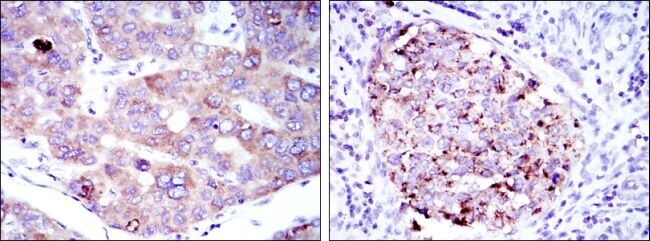
- Immunohistochemical analysis of paraffin-embedded liver cancer tissues (left) and kidney cancer tissues (right) using EEF2 monoclonal antibody (Product # MA5-15833) followed with DAB staining.
- Submitted by
- Invitrogen Antibodies (provider)
- Main image

- Experimental details
- Immunohistochemical analysis of paraffin-embedded prostate cancer tissues (left) and tonsil tissues (right) using EEF2 monoclonal antibody (Product # MA5-15833) followed with DAB staining.
