 Explore
 Validate
 Learn
Learn Immunocytochemistry
 Immunohistochemistry
Antibody data
- Antibody Data
- Antigen structure
- References [7]
- Comments [0]
- Validations
- Immunocytochemistry [1]
Submit
Validation data
Reference
Comment
Report error
- Product number
- HPA043785 - Provider product page

- Provider
- Atlas Antibodies
- Proper citation
- Atlas Antibodies Cat#HPA043785, RRID:AB_10961859
- Product name
- Anti-SLC38A9
- Antibody type
- Polyclonal
- Description
- Polyclonal Antibody against Human SLC38A9, Gene description: solute carrier family 38, member 9, Alternative Gene Names: FLJ90709, Validated applications: IHC, ICC, Uniprot ID: Q8NBW4, Storage: Store at +4°C for short term storage. Long time storage is recommended at -20°C.
- Reactivity
- Human
- Host
- Rabbit
- Conjugate
- Unconjugated
- Isotype
- IgG
- Vial size
- 100 µl
- Concentration
- 0.2 mg/ml
- Storage
- Store at +4°C for short term storage. Long time storage is recommended at -20°C.
- Handling
- The antibody solution should be gently mixed before use.
Submitted references Amino Acid-Dependent mTORC1 Regulation by the Lysosomal Membrane Protein SLC38A9
NRF3 activates mTORC1 arginine-dependently for cancer cell viability
TASL is the SLC15A4-associated adaptor for IRF5 activation by TLR7–9
Galectins Control mTOR in Response to Endomembrane Damage
TFEB-driven endocytosis coordinates MTORC1 signaling and autophagy
The gene expression of the neuronal protein, SLC38A9, changes in mouse brain after in vivo starvation and high-fat diet
SLC38A9 is a component of the lysosomal amino acid sensing machinery that controls mTORC1
Jung J, Genau H, Behrends C
Molecular and Cellular Biology 2023;35(14):2479-2494
Molecular and Cellular Biology 2023;35(14):2479-2494
NRF3 activates mTORC1 arginine-dependently for cancer cell viability
Hirose S, Waku T, Tani M, Masuda H, Endo K, Ashitani S, Aketa I, Kitano H, Nakada S, Wada A, Hatanaka A, Osawa T, Soga T, Kobayashi A
iScience 2023;26(2):106045
iScience 2023;26(2):106045
TASL is the SLC15A4-associated adaptor for IRF5 activation by TLR7–9
Heinz L, Lee J, Kapoor U, Kartnig F, Sedlyarov V, Papakostas K, César-Razquin A, Essletzbichler P, Goldmann U, Stefanovic A, Bigenzahn J, Scorzoni S, Pizzagalli M, Bensimon A, Müller A, King F, Li J, Girardi E, Mbow M, Whitehurst C, Rebsamen M, Superti-Furga G
Nature 2020;581(7808):316-322
Nature 2020;581(7808):316-322
Galectins Control mTOR in Response to Endomembrane Damage
Jia J, Abudu Y, Claude-Taupin A, Gu Y, Kumar S, Choi S, Peters R, Mudd M, Allers L, Salemi M, Phinney B, Johansen T, Deretic V
Molecular Cell 2018;70(1):120-135.e8
Molecular Cell 2018;70(1):120-135.e8
TFEB-driven endocytosis coordinates MTORC1 signaling and autophagy
Nnah I, Wang B, Saqcena C, Weber G, Bonder E, Bagley D, De Cegli R, Napolitano G, Medina D, Ballabio A, Dobrowolski R
Autophagy 2018;15(1):151-164
Autophagy 2018;15(1):151-164
The gene expression of the neuronal protein, SLC38A9, changes in mouse brain after in vivo starvation and high-fat diet
Jadhao S, Hellsten S, Eriksson M, Lekholm E, Arapi V, Perland E, Fredriksson R
PLOS ONE 2017;12(2):e0172917
PLOS ONE 2017;12(2):e0172917
SLC38A9 is a component of the lysosomal amino acid sensing machinery that controls mTORC1
Rebsamen M, Pochini L, Stasyk T, de Araújo M, Galluccio M, Kandasamy R, Snijder B, Fauster A, Rudashevskaya E, Bruckner M, Scorzoni S, Filipek P, Huber K, Bigenzahn J, Heinz L, Kraft C, Bennett K, Indiveri C, Huber L, Superti-Furga G
Nature 2015;519(7544):477-481
Nature 2015;519(7544):477-481
No comments: Submit comment
Supportive validation
- Submitted by
- Atlas Antibodies (provider)
- Main image
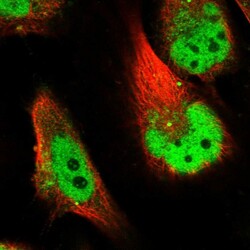
- Experimental details
- Immunofluorescent staining of human cell line U-251 MG shows localization to nucleoplasm & vesicles.
- Sample type
- Human
