Explore
 Validate
 Learn
Learn Western blot
 Immunocytochemistry
Antibody data
- Antibody Data
- Antigen structure
- References [2]
- Comments [0]
- Validations
- Immunocytochemistry [2]
- Immunohistochemistry [1]
- Flow cytometry [1]
- Other assay [1]
Submit
Validation data
Reference
Comment
Report error
- Product number
- MA5-15565 - Provider product page

- Provider
- Invitrogen Antibodies
- Product name
- DDX4 Monoclonal Antibody (2F9H5)
- Antibody type
- Monoclonal
- Antigen
- Purifed from natural sources
- Description
- MA5-15565 targets DDX4 in FACS, IF, IHC, and WB applications and shows reactivity with Human samples. The MA5-15565 immunogen is purified recombinant fragment of human DDX4 expressed in E. Coli. MA5-15565 detects DDX4 which has a predicted molecular weight of approximately 76kDa.
- Reactivity
- Human, Mouse
- Host
- Mouse
- Isotype
- IgG
- Antibody clone number
- 2F9H5
- Vial size
- 100 μg
- Concentration
- 1 mg/mL
- Storage
- Store at 4°C short term. For long term storage, store at -20°C, avoiding freeze/thaw cycles.
Submitted references "Mitotic Slippage" and Extranuclear DNA in Cancer Chemoresistance: A Focus on Telomeres.
Nuclear Factor Y (NF-Y) Modulates Encystation in Entamoeba via Stage-Specific Expression of the NF-YB and NF-YC Subunits.
Salmina K, Bojko A, Inashkina I, Staniak K, Dudkowska M, Podlesniy P, Rumnieks F, Vainshelbaum NM, Pjanova D, Sikora E, Erenpreisa J
International journal of molecular sciences 2020 Apr 16;21(8)
International journal of molecular sciences 2020 Apr 16;21(8)
Nuclear Factor Y (NF-Y) Modulates Encystation in Entamoeba via Stage-Specific Expression of the NF-YB and NF-YC Subunits.
Manna D, Singh U
mBio 2019 Jun 18;10(3)
mBio 2019 Jun 18;10(3)
No comments: Submit comment
Supportive validation
- Submitted by
- Invitrogen Antibodies (provider)
- Main image

- Experimental details
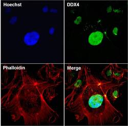
- Immunofluorescent analysis of DDX4 (green) in NTERA-2 cells. The cells were fixed with 4% paraformaldehyde for 15 minutes at -20°C, permeabilized with 0.1% Triton X-100 for 15 minutes, and blocked with 3% BSA for 30 minutes at room temperature. Cells were stained with a DDX4mouse monoclonal antibody (Product # MA5-15565) at a concentration of 5 µg/mL in blocking buffer for 1 hour at room temperature, and then incubated with a Goat anti-Mouse IgG (H+L) Secondary Antibody, Alexa Fluor Plus 488 conjugate (Product # A32731) at a dilution of 1:500 for at least 30 minutes at a room temperature in the dark (green). F-actin (red) was stained with Dylight 554 Phalloidin. Nuclei (blue) were stained with Hoechst 33342 (Product # 62249). Images were taken on a Thermo Scientific ToxInsight Instrument at 20X magnification.

- Submitted by
- Invitrogen Antibodies (provider)
- Main image
- Experimental details
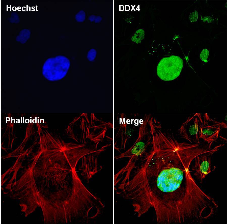
- Immunofluorescent analysis of DDX4 (green) in NTERA-2 cells. The cells were fixed with 4% paraformaldehyde for 15 minutes at -20°C, permeabilized with 0.1% Triton X-100 for 15 minutes, and blocked with 3% BSA for 30 minutes at room temperature. Cells were stained with a DDX4mouse monoclonal antibody (Product # MA5-15565) at a concentration of 5 µg/mL in blocking buffer for 1 hour at room temperature, and then incubated with a Goat anti-Mouse IgG (H+L) Secondary Antibody, Alexa Fluor Plus 488 conjugate (Product # A32731) at a dilution of 1:500 for at least 30 minutes at a room temperature in the dark (green). F-actin (red) was stained with Dylight 554 Phalloidin. Nuclei (blue) were stained with Hoechst 33342 (Product # 62249). Images were taken on a Thermo Scientific ToxInsight Instrument at 20X magnification.
Supportive validation
- Submitted by
- Invitrogen Antibodies (provider)
- Main image
- Experimental details
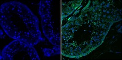
- Immunofluorescent analysis of DDX4 (green) on Human Testis tissue with a DDX4 mouse monoclonal antibody (Product # MA5-15565, right panel) at a concentration of 5 µg/mL in blocking buffer for 1 hour at room temperature compared with negative control in the absence of the primary antibody (left panel), and then incubated with a Goat anti-Mouse IgG (H+L) Secondary Antibody, DyLight 488 conjugate at a dilution of 1:500 for at least 30 minutes at a room temperature in the dark (green). Nuclei (blue) were stained with DAPI. Images were taken at 60X magnification.

Supportive validation
- Submitted by
- Invitrogen Antibodies (provider)
- Main image

- Experimental details
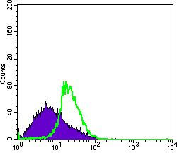
- Flow cytometric analysis of MSCS cells using DDX4 monoclonal antibody (Product # MA5-15565) (green) and negative control (purple).
Supportive validation
- Submitted by
- Invitrogen Antibodies (provider)
- Main image
- Experimental details
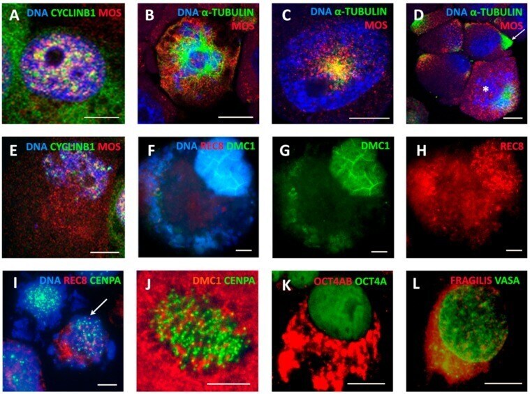
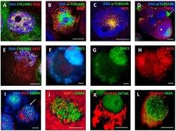
- Figure 8 Expression of meiotic and germline proteins in MS and giant cells found by Immunofluorescence--representative images for at least three experiments: ( A ) a tetraploid cell nucleus enriched with MOS-kinase (sc-86) colocalized and juxtaposed with CYCLIN B1 (DOX-D2); ( B ) an attachment of MOS to the centrosomes and microtubules of the tripolar mitosis (DOX-D4); ( C ) MOS and alpha-TUBULIN form a monopolar spindle in the early prophase (DOX-D4); ( D ) MOS is attached to interphase centrosomes (arrow) and shows a remnant of a monopolar spindle in MS (asterisk) []; ( E ) the restituting nucleus in MS becomes poor with MOS and CYCLIN B1 (DOX-D4); ( F - H ) a giant cell in MS releasing cytoplasmic DNA shows the enrichment of the cell nucleus with DMC1 (meiotic recombinase) and REC8 (meiotic cohesin) (DOX-D19); ( I ) REC8 grains are scarcely inserted in the kinetochore chains in the MS cell (arrow) (DOX-D4); ( J ) DMC1 grains are scarcely inserted in the MS cell (DOX-D7; []); ( K ) a giant cell enriched with OCT4A in the cell nucleus (a monoclonal Ab) and OCT4B in the cytoplasm (DOX-D5); and ( L ) a giant cell enriched with the germ markers, DDX4/VASA in the cell nucleus and FRAGILIS in the cytoplasm (DOX-D7). Bars = 10 um.