 Explore
 Validate
 Learn
Learn Western blot
Antibody data
- Antibody Data
- Antigen structure
- References [0]
- Comments [0]
- Validations
- Western blot [3]
- Immunocytochemistry [3]
Submit
Validation data
Reference
Comment
Report error
- Product number
- PA1-32336 - Provider product page

- Provider
- Invitrogen Antibodies
- Product name
- NTHL1 Polyclonal Antibody
- Antibody type
- Polyclonal
- Antigen
- Recombinant full-length protein
- Description
- Description: The monoclonal antibody 2D3 reacts to CD47 also known as integrin-associated protein (IAP), and neurophilin. CD47 is a glycosylated five transmembrane protein with a small alternatively spliced cytoplasmic domain. CD47 is involved in adhesion through interactions with SIRP (signal regulator protein) and is non-covalently associated with beta3 integrins CD51/CD61 and CD41/CD61. Furthermore this interaction can mediate bi-directional signaling to modify neural synaptic activity and regulate the phagocytic activities of macrophages. CD47 is the receptor for thrombospondin. T cell expression of CD47 can mediate activation or apoptosis (in the presence of high levels of thrombospondin). Expression is found in the majority of hematopoietic cells including T and B cells, monocytes, platelets and erythrocytes (as part of the Rh complex). Expression is also found in non-hematopoietic cells.
- Reactivity
- Human, Mouse, Rat
- Host
- Rabbit
- Isotype
- IgG
- Vial size
- 100 µL
- Concentration
- Conc. Not Determined
- Storage
- Store at 4°C short term. For long term storage, store at -20°C, avoiding freeze/thaw cycles.
No comments: Submit comment
Supportive validation
- Submitted by
- Invitrogen Antibodies (provider)
- Main image

- Experimental details
- Western blot of NTH in A549 cells using a NTH polyclonal antibody (Product # PA1-32336). A band is seen at 35 kDa representing NTH1.

- Submitted by
- Invitrogen Antibodies (provider)
- Main image

- Experimental details
- Western Blot analysis of A549 cell lysate using NTHL1 Polyclonal Antibody (Product # PA1-32336).

- Submitted by
- Invitrogen Antibodies (provider)
- Main image

- Experimental details
- Western blot was performed using Anti-NTHL1 Polyclonal Antibody (Product # PA1-32336) and a 34 kDa band corresponding to Endonuclease III-like protein 1 was observed across the cell lines tested along with an uncharacterised band (*) at ~50 kDa and increased upon serum release for 24 Hours followed by serum starvation for 72 Hours in HaCa T. Whole cell extracts (30 µg lysate) of HeLa (Lane 1), Hep G2 (Lane 2), Reh(Lane 3), MCF7 (Lane 4), A-431 (Lane 5), HaCa T serum starved for 72 Hours (Lane 6) and serum starvation for 72 Hours followed by Serum release for 24 Hours (Lane 7) were electrophoresed using NuPAGE™ 4-12% Bis-Tris Protein Gel (Product # NP0321BOX). Resolved proteins were then transferred onto a Nitrocellulose membrane (Product # LC2001) by iBlot® 2 Dry Blotting System (Product # IB21001). The blot was probed with the primary antibody (1:1000 dilution) and detected by chemiluminescence with Goat anti-Rabbit IgG (H+L) Superclonal™ Recombinant Secondary Antibody, HRP (Product # A27036, 1:4000 dilution) using the iBright FL 1000 (Product # A32752). Chemiluminescent detection was performed using Novex® ECL Chemiluminescent Substrate Reagent Kit (Product # WP20005).

Supportive validation
- Submitted by
- Invitrogen Antibodies (provider)
- Main image

- Experimental details
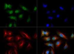
- Immunofluorescent analysis of NTH in Hela cells using a NTH polyclonal antibody (Product # PA1-32336) at a dilution of 1:500 and a Dylight 488 secondary antibody (Green). Alpha-tubulin and nuclei were counterstained using Dylight 550 (Red) and DAPI (Blue).

- Submitted by
- Invitrogen Antibodies (provider)
- Main image
- Experimental details
- Immunocytochemistry-Immunofluorescence analysis of NTHL1 in HeLa cells using NTHL1 Polyclonal Antibody (Product # PA1-32336) (Green) at a dilution of 1:500. Red : Tubulin. Blue: DAPI.

- Submitted by
- Invitrogen Antibodies (provider)
- Main image
- Experimental details
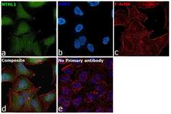
- Immunofluorescence analysis of Endonuclease III-like protein 1 was performed using 70% confluent log phase HeLa cells. The cells were fixed with 4% paraformaldehyde for 10 minutes, permeabilized with 0.1% Triton™ X-100 for 10 minutes, and blocked with 2% BSA for 45 minutes at room temperature. The cells were labeled with NTHL1 Polyclonal Antibody (Product # PA1-32336) at 1:100 dilution in 0.1% BSA, incubated at 4 degree celsius overnight and then labeled with Goat anti-Rabbit IgG (H+L) Superclonal™ Recombinant Secondary Antibody, Alexa Fluor® 488 conjugate (Product # A27034), (1:2000 dilution), for 45 minutes at room temperature (Panel a: Green). Nuclei (Panel b:Blue) were stained with ProLong™ Diamond Antifade Mountant with DAPI (Product # P36962). F-actin (Panel c: Red) was stained with Rhodamine Phalloidin (Product # R415, 1:300 dilution). Panel d represents the merged image showing nuclear and mitochondrial localization. Panel e represents control cells with no primary antibody to assess background. The images were captured at 60X magnification.
