 Explore
 Validate
 Learn
Learn Western blot
 Immunocytochemistry
Antibody data
- Antibody Data
- Antigen structure
- References [9]
- Comments [0]
- Validations
- Western blot [2]
- Immunohistochemistry [5]
Submit
Validation data
Reference
Comment
Report error
- Product number
- NB100-108 - Provider product page

- Provider
- Novus Biologicals
- Proper citation
- Novus Cat#NB100-108, RRID:AB_10080489
- Product name
- Rabbit Polyclonal NTH1 Antibody
- Antibody type
- Polyclonal
- Description
- Immunogen affinity purified.
- Reactivity
- Human, Mouse, Rat
- Host
- Rabbit
- Isotype
- IgG
- Vial size
- 0.2 ml
- Concentration
- 1.1 mg/ml
- Storage
- Aliquot and store at -20C or -80C. Avoid freeze-thaw cycles.
Submitted references Overexpression of the base excision repair NTHL1 glycosylase causes genomic instability and early cellular hallmarks of cancer.
Induction of base excision repair enzymes NTH1 and APE1 in rat spleen following aniline exposure.
Poor maternal nutrition followed by accelerated postnatal growth leads to alterations in DNA damage and repair, oxidative and nitrosative stress, and oxidative defense capacity in rat heart.
Altered expression of the human base excision repair gene NTH1 in gastric cancer.
S1P(5) is required for sphingosine 1-phosphate-induced autophagy in human prostate cancer PC-3 cells.
p21(Cip1/WAF1/Sdi1) does not affect expression of base excision DNA repair enzymes during chronic oxidative stress.
Neuronal expression of caspase-1 immunoreactivity in the rat central nervous system.
Neuronal expression of caspase-1 immunoreactivity in the rat central nervous system.
Zinc(II) inhibits the release of thyroid and glucocorticoid receptors from chromatin of cultured GC cells.
Limpose KL, Trego KS, Li Z, Leung SW, Sarker AH, Shah JA, Ramalingam SS, Werner EM, Dynan WS, Cooper PK, Corbett AH, Doetsch PW
Nucleic acids research 2018 May 18;46(9):4515-4532
Nucleic acids research 2018 May 18;46(9):4515-4532
Induction of base excision repair enzymes NTH1 and APE1 in rat spleen following aniline exposure.
Ma H, Wang J, Abdel-Rahman SZ, Boor PJ, Khan MF
Toxicology and applied pharmacology 2013 Mar 15;267(3):276-83
Toxicology and applied pharmacology 2013 Mar 15;267(3):276-83
Poor maternal nutrition followed by accelerated postnatal growth leads to alterations in DNA damage and repair, oxidative and nitrosative stress, and oxidative defense capacity in rat heart.
Tarry-Adkins JL, Martin-Gronert MS, Fernandez-Twinn DS, Hargreaves I, Alfaradhi MZ, Land JM, Aiken CE, Ozanne SE
FASEB journal : official publication of the Federation of American Societies for Experimental Biology 2013 Jan;27(1):379-90
FASEB journal : official publication of the Federation of American Societies for Experimental Biology 2013 Jan;27(1):379-90
Altered expression of the human base excision repair gene NTH1 in gastric cancer.
Goto M, Shinmura K, Igarashi H, Kobayashi M, Konno H, Yamada H, Iwaizumi M, Kageyama S, Tsuneyoshi T, Tsugane S, Sugimura H
Carcinogenesis 2009 Aug;30(8):1345-52
Carcinogenesis 2009 Aug;30(8):1345-52
S1P(5) is required for sphingosine 1-phosphate-induced autophagy in human prostate cancer PC-3 cells.
Chang CL, Ho MC, Lee PH, Hsu CY, Huang WP, Lee H
American journal of physiology. Cell physiology 2009 Aug;297(2):C451-8
American journal of physiology. Cell physiology 2009 Aug;297(2):C451-8
p21(Cip1/WAF1/Sdi1) does not affect expression of base excision DNA repair enzymes during chronic oxidative stress.
O'reilly MA, Vitiello PF, Gehen SC, Staversky RJ
Antioxidants & redox signaling 2005 May-Jun;7(5-6):719-25
Antioxidants & redox signaling 2005 May-Jun;7(5-6):719-25
Neuronal expression of caspase-1 immunoreactivity in the rat central nervous system.
Lindberg C, Eriksson C, Van Dam AM, Winblad B, Schultzberg M
Journal of neuroimmunology 2004 Jan;146(1-2):99-113
Journal of neuroimmunology 2004 Jan;146(1-2):99-113
Neuronal expression of caspase-1 immunoreactivity in the rat central nervous system.
Lindberg C, Eriksson C, Van Dam AM, Winblad B, Schultzberg M
Journal of neuroimmunology 2004 Jan;146(1-2):99-113
Journal of neuroimmunology 2004 Jan;146(1-2):99-113
Zinc(II) inhibits the release of thyroid and glucocorticoid receptors from chromatin of cultured GC cells.
Ramirez IJ, Halwer M, Shapiro LE, Surks MI
Hormone and metabolic research = Hormon- und Stoffwechselforschung = Hormones et metabolisme 1991 Apr;23(4):155-61
Hormone and metabolic research = Hormon- und Stoffwechselforschung = Hormones et metabolisme 1991 Apr;23(4):155-61
No comments: Submit comment
Supportive validation
- Submitted by
- Novus Biologicals (provider)
- Main image
- Experimental details
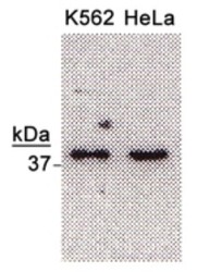
- Western Blot: NTH1 Antibody [NB100-108] - Detection of NTH1 (37 kDa) from Hela & K562 cell extracts using NB100-108 (1:500). WB data courtesy of Mark Kelly, Indiana Univ.

- Submitted by
- Novus Biologicals (provider)
- Main image
- Experimental details
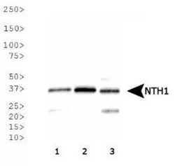
- Western Blot: NTH1 Antibody [NB100-108] - Analysis of NTH1 expression in 1) HeLa, 2) A-431, 3) MCF7 whole cell lysates.
Supportive validation
- Submitted by
- Novus Biologicals (provider)
- Main image

- Experimental details
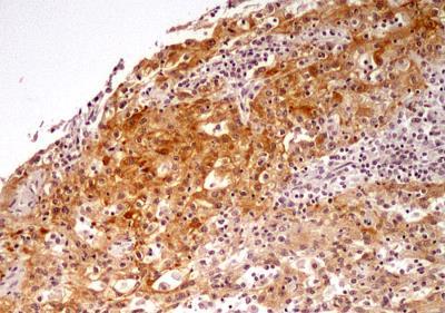
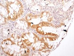
- Immunohistochemistry-Paraffin: NTH1 Antibody [NB100-108] - IHC analysis of formalin-fixed paraffin-embedded tissue section of human kidney cancer using 5 ug/ml concentration of NTH1 antibody. The renal cancer cells showed nuclear and cytoplasmic positivity for NTH1 protein, whereas, the tumor stroma was largely negative for immunostaining.
- Submitted by
- Novus Biologicals (provider)
- Main image
- Experimental details
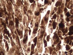
- Immunohistochemistry-Paraffin: NTH1 Antibody [NB100-108] - IHC analysis of formalin-fixed paraffin-embedded tissue section of human lymph node cancer using 5 ug/ml concentration of NTH1 antibody. The representative image shows strong nuclear and cytoplasmic positivity of NTH1 protein in the lymph node cancer cells.
- Submitted by
- Novus Biologicals (provider)
- Main image
- Experimental details
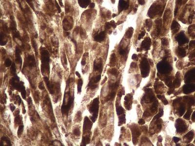
- Immunohistochemistry-Paraffin: NTH1 Antibody [NB100-108] - IHC analysis of formalin-fixed paraffin-embedded tissue section of human rectal cancer using 5 ug/ml concentration of NTH1 antibody. The rectal cancer cells as well as the goblet cells in glandular areas showed nuclear-cytoplasmic positivity for NTH1 protein. The cells of tumor stroma did not develop immunostaining for this protein.

- Submitted by
- Novus Biologicals (provider)
- Main image
- Experimental details
- Immunohistochemistry-Paraffin: NTH1 Antibody [NB100-108] - IHC analysis of formalin-fixed paraffin-embedded tissue section of human rectal cancer using 5 ug/ml concentration of NTH1 antibody. This representative image at high resolution depicts nuclear-cytoplasmic positivity for NTH1 protein in rectal cancer cells and the goblet cells. The cells of tumor stroma did not develop any immunostaining for this protein.

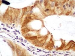
- Submitted by
- Novus Biologicals (provider)
- Main image
- Experimental details
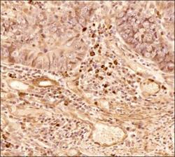
- Immunohistochemistry-Paraffin: NTH1 Antibody [NB100-108] - IHC analysis of a formalin fixed paraffin embedded (FFPE) tissue section of human colon adenocarcinoma using NTH1 antibody at 5ug/ml concentration (1:200 dilution). The primary antibody binding to NTH1/ NTHL1 antigen was detected using HRP conjugated anti-rabbit secondary antibody with DAB reagent, and the sections were further counterstained with hematoxylin for labeling cellular nuclei. The NTH1 antibody generated an expected nuclear cytoplasmic staining of NTH1 protein in colon cancer cells as well as the cells of tumor stroma including cancer associated fibroblasts. The nuclear staining of NTH1 was very strong in a sub-set of colon cancer cells.
