Explore
 Validate
 Learn
Learn Western blot
Antibody data
- Antibody Data
- Antigen structure
- References [0]
- Comments [0]
- Validations
- Western blot [1]
- Immunocytochemistry [2]
Submit
Validation data
Reference
Comment
Report error
- Product number
- PA5-85179 - Provider product page

- Provider
- Invitrogen Antibodies
- Product name
- NTHL1 Polyclonal Antibody
- Antibody type
- Polyclonal
- Antigen
- Recombinant full-length protein
- Description
- Keep as concentrated solution.
- Concentration
- 0.78 mg/mL
No comments: Submit comment
Supportive validation
- Submitted by
- Invitrogen Antibodies (provider)
- Main image

- Experimental details
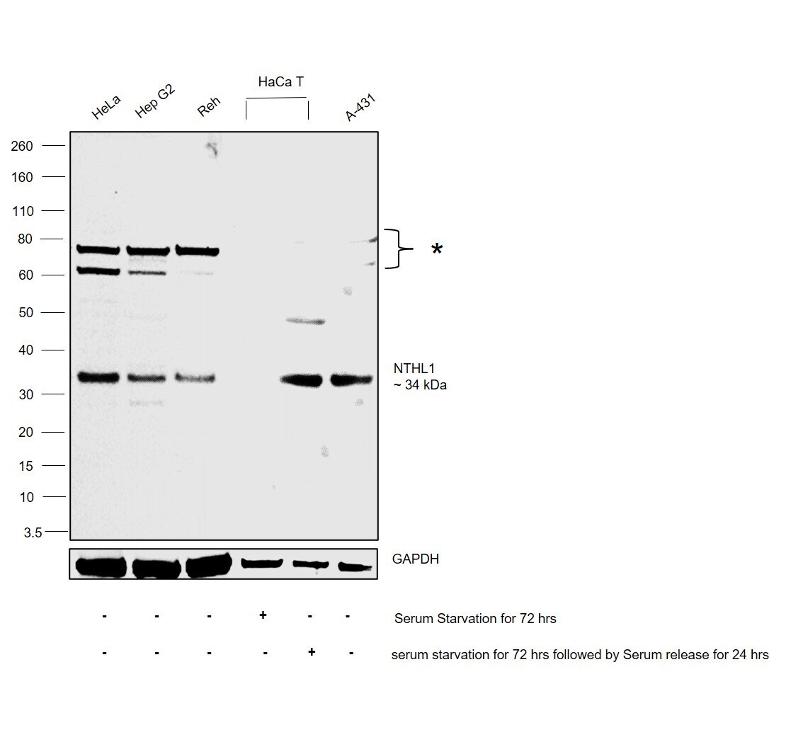
- Western blot was performed using Anti-NTHL1 Polyclonal Antibody (Product # PA5-85179) and a 34 kDa band corresponding to Endonuclease III-like protein 1 was observed along with uncharacterised bands (*) at ~65-75 kDa across the cell lines tested and increased upon serum starvation for 72 Hours followed by Serum release for 24 Hours in HaCa T. Whole cell extracts (30 µg lysate) of HeLa (Lane 1), Hep G2 (Lane 2), Reh (Lane 3), HaCa T serum starvated for 72 Hours (Lane 4), HaCa T serum starvation for 72 Hours followed by serum release for 24 Hours (Lane 5) and A-431 (Lane 6) were electrophoresed using NuPAGE™ 4-12% Bis-Tris Protein Gel (Product # NP0321BOX). Resolved proteins were then transferred onto a PVDF membrane (Product # LC2001) by iBlot® 2 Dry Blotting System (Product # IB21001). The blot was probed with the primary antibody (1:1,000 dilution) and detected by chemiluminescence with Goat anti-Rabbit IgG (H+L) Superclonal™ Recombinant Secondary Antibody, HRP (Product # A27036, 1:4,000 dilution) using the iBright FL 1000 (Product # A32752). Chemiluminescent detection was performed using Novex® ECL Chemiluminescent Substrate Reagent Kit (Product # WP20005).
Supportive validation
- Submitted by
- Invitrogen Antibodies (provider)
- Main image

- Experimental details
- NTHL1 Polyclonal Antibody detects NTH1 protein at nucleus by immunofluorescent analysis. Sample: A549 cells were fixed in 4% paraformaldehyde at RT for 15 min. Green: NTH1 protein stained by NTHL1 Polyclonal Antibody (Product # PA5-85179) diluted at 1:1,000. Blue: Hoechst 33342 staining.

- Submitted by
- Invitrogen Antibodies (provider)
- Main image
- Experimental details
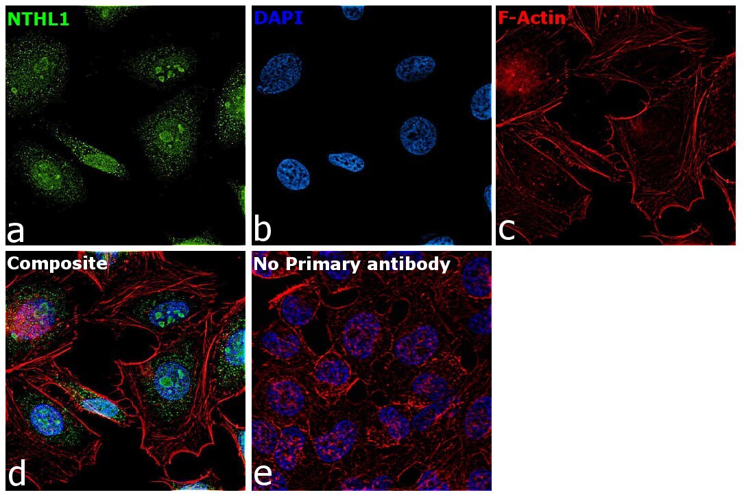
- Immunofluorescence analysis of Endonuclease III-like protein 1 was performed using 70% confluent log phase HeLa cells. The cells were fixed with 4% paraformaldehyde for 15 minutes, permeabilized with 0.1% Triton™ X-100 for 10 minutes, and blocked with 2% BSA for 45 minutes at room temperature. The cells were labeled with NTHL1 Polyclonal Antibody (Product # PA5-85179) at 1:100 dilution in 0.1% BSA, incubated at 4 degree celsius overnight and then labeled with Goat anti-Rabbit IgG (H+L) Superclonal™ Recombinant Secondary Antibody, Alexa Fluor® 488 conjugate (Product # A27034), (1:2000 dilution), for 45 minutes at room temperature (Panel a: Green). Nuclei (Panel b:Blue) were stained with ProLong™ Diamond Antifade Mountant with DAPI (Product # P36962). F-actin (Panel c: Red) was stained with Rhodamine Phalloidin (Product # R415, 1:300 dilution). Panel d represents the merged image showing nuclear and mitochondrial localization. Panel e represents control cells with no primary antibody to assess background. The images were captured at 60X magnification.