Explore
 Validate
 Learn
Learn Western blot
Antibody data
- Antibody Data
- Antigen structure
- References [3]
- Comments [0]
- Validations
- Western blot [1]
- Immunocytochemistry [1]
Submit
Validation data
Reference
Comment
Report error
- Product number
- AF3666 - Provider product page

- Provider
- R&D Systems
- Product name
- Human DSCAM Long Isoform Antibody
- Antibody type
- Polyclonal
- Description
- Antigen Affinity-purified. Detects human DSCAM in direct ELISAs and Western blots. In direct ELISAs, less than 10% cross-reactivity with recombinant human DSCAM-L1 is observed.
- Reactivity
- Human
- Host
- Goat
- Conjugate
- Unconjugated
- Antigen sequence
O60469- Isotype
- IgG
- Vial size
- 100 ug
- Concentration
- LYOPH
- Storage
- Use a manual defrost freezer and avoid repeated freeze-thaw cycles. 12 months from date of receipt, -20 to -70 °C as supplied. 1 month, 2 to 8 °C under sterile conditions after reconstitution. 6 months, -20 to -70 °C under sterile conditions after reconstitution.
Submitted references DSCAM promotes axon fasciculation and growth in the developing optic pathway.
Replacing the PDZ-interacting C-termini of DSCAM and DSCAML1 with epitope tags causes different phenotypic severity in different cell populations.
Neurite arborization and mosaic spacing in the mouse retina require DSCAM.
Bruce FM, Brown S, Smith JN, Fuerst PG, Erskine L
Proceedings of the National Academy of Sciences of the United States of America 2017 Feb 14;114(7):1702-1707
Proceedings of the National Academy of Sciences of the United States of America 2017 Feb 14;114(7):1702-1707
Replacing the PDZ-interacting C-termini of DSCAM and DSCAML1 with epitope tags causes different phenotypic severity in different cell populations.
Garrett AM, Tadenev AL, Hammond YT, Fuerst PG, Burgess RW
eLife 2016 Sep 16;5
eLife 2016 Sep 16;5
Neurite arborization and mosaic spacing in the mouse retina require DSCAM.
Fuerst PG, Koizumi A, Masland RH, Burgess RW
Nature 2008 Jan 24;451(7177):470-4
Nature 2008 Jan 24;451(7177):470-4
No comments: Submit comment
Supportive validation
- Submitted by
- R&D Systems (provider)
- Main image

- Experimental details
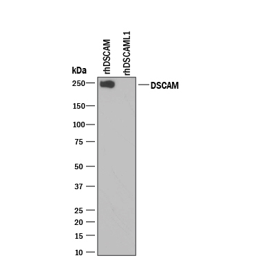
- Detection of Recombinant Human DSCAM by Western Blot. Western blot shows 25 ng of Recombinant Human DSCAM (Catalog # 3666-DS) and Recombinant Human DSCAM-L1 (Catalog # 3315-DL). PVDF Membrane was probed with 0.1 µg/mL of Goat Anti-Human DSCAM Long Isoform Antigen Affinity-purified Polyclonal Antibody (Catalog # AF3666) followed by HRP-conjugated Anti-Goat IgG Secondary Antibody (Catalog # HAF109). A specific band was detected for DSCAM at approximately 250 kDa (as indicated). This experiment was conducted under reducing conditions and using Immunoblot Buffer Group 3.
Supportive validation
- Submitted by
- R&D Systems (provider)
- Main image

- Experimental details
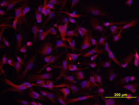
- DSCAM in A172 Human Cell Line. DSCAM was detected in immersion fixed A172 human glioblastoma cell line using 10 µg/mL Goat Anti-Human DSCAM Long Isoform Antigen Affinity-purified Polyclonal Antibody (Catalog # AF3666) for 3 hours at room temperature. Cells were stained with the NorthernLights™ 557-conjugated Anti-Goat IgG Secondary Antibody (red; Catalog # NL001) and counterstained with DAPI (blue). View our protocol for Fluorescent ICC Staining of Cells on Coverslips.