Explore
 Validate
 Learn
Learn Western blot
Antibody data
- Antibody Data
- Antigen structure
- References [1]
- Comments [0]
- Validations
- Western blot [3]
- Immunocytochemistry [1]
- Flow cytometry [1]
Submit
Validation data
Reference
Comment
Report error
- Product number
- PA5-17444 - Provider product page

- Provider
- Invitrogen Antibodies
- Product name
- Profilin 1 Polyclonal Antibody
- Antibody type
- Polyclonal
- Antigen
- Synthetic peptide
- Description
- It is not recommended to aliquot this antibody.
- Reactivity
- Human, Mouse, Rat, Bovine
- Host
- Rabbit
- Isotype
- IgG
- Vial size
- 100 µL
- Concentration
- 78 µg/mL
- Storage
- -20°C
Submitted references Profilin 1 delivery tunes cytoskeletal dynamics toward CNS axon regeneration.
Pinto-Costa R, Sousa SC, Leite SC, Nogueira-Rodrigues J, Ferreira da Silva T, Machado D, Marques J, Costa AC, Liz MA, Bartolini F, Brites P, Costell M, Fässler R, Sousa MM
The Journal of clinical investigation 2020 Apr 1;130(4):2024-2040
The Journal of clinical investigation 2020 Apr 1;130(4):2024-2040
No comments: Submit comment
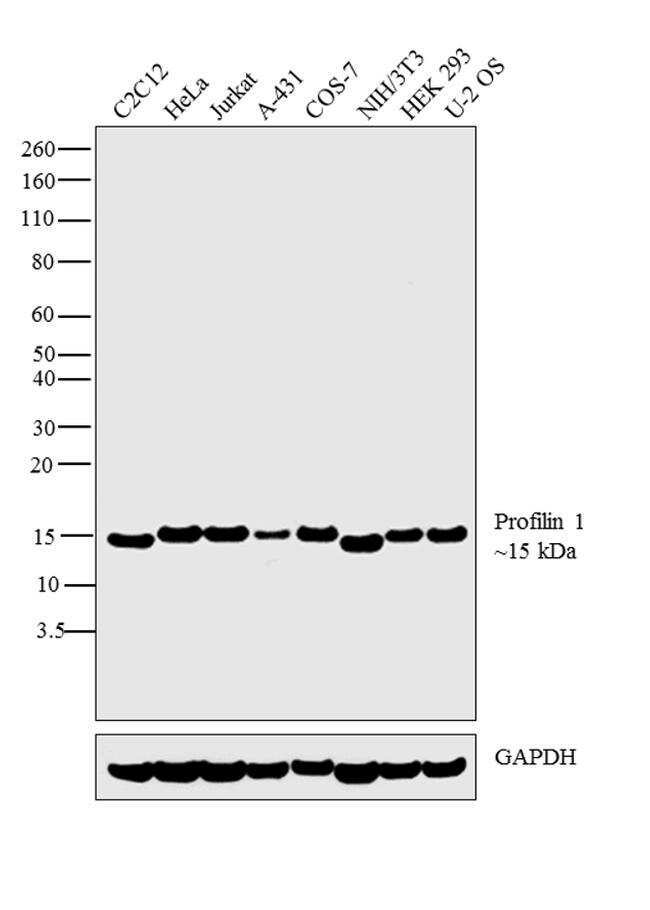
Supportive validation
- Submitted by
- Invitrogen Antibodies (provider)
- Main image

- Experimental details
- Western blot analysis was performed whole cell extracts (30 µg lysate) of C2C12 (Lane 1), HeLa (Lane 2), Jurkat (Lane 3), A-431 (Lane 4), COS-7 (Lane 5), NIH/3T3 (Lane 6), HEK 293 (Lane 7) and U-2 OS (Lane 8). The blot was probed with Anti-Profilin 1 antibody (Product # PA5-17444, 1:500) and detected by chemiluminescence using Goat anti-Rabbit IgG (H+L) Superclonal™ Secondary Antibody, HRP conjugate (Product # A27036, 0.25 µg/mL, 1:4000 dilution). A 15 kDa band corresponding to Profilin 1 was observed across the cell lines tested.
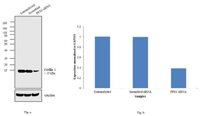
- Submitted by
- Invitrogen Antibodies (provider)
- Main image
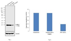
- Experimental details
- Knockdown of Profilin 1 was achieved by transfecting HeLa cells with Profilin 1 specific siRNAs (Silencer® select Product # s10375, s10376). Western blot analysis (Fig. a) was performed using whole cell extracts from the Profilin 1 knockdown cells (lane 3), non-specific scrambled siRNA transfected cells (lane 2) and untransfected cells (lane 1). The blots were probed with Profilin 1 Polyclonal Antibody (Product # PA5-17444, 1:1000 dilution) and Goat anti-Rabbit IgG (H+L) Superclonal™ Secondary Antibody, HRP conjugate (Product # A27036, 0.25 µg/mL, 1:4000 dilution). Densitometric analysis of this western blot is shown in histogram (Fig. b). Decrease in signal upon siRNA mediated knock down confirms that antibody is specific to Profilin 1.
- Submitted by
- Invitrogen Antibodies (provider)
- Main image

- Experimental details
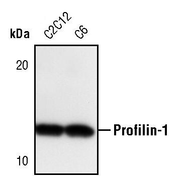
- Western blot analysis of Profilin-1 in extracts from C2C12 and C6 cell lines using Profilin-1 polyclonal antibody (Product # PA5-17444).
Supportive validation
- Submitted by
- Invitrogen Antibodies (provider)
- Main image

- Experimental details
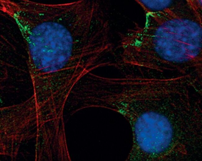
- Immunofluorescent analysis of Profilin-1 in C2C12 cells using a Profilin-1 polyclonal antibody (Product # PA5-17444) (green). Actin filaments are labeled with a fluorescent red phalloidin. DNA is labeled using a fluorescent blue dye.
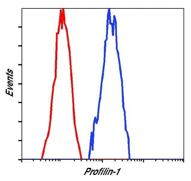
Supportive validation
- Submitted by
- Invitrogen Antibodies (provider)
- Main image
- Experimental details
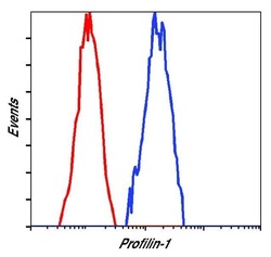
- Flow cytometric analysis of Profilin-1 in HeLa cells using a Profilin-1 polyclonal antibody (Product # PA5-17444) (blue) compared to a nonspecific negative control antibody (red).