Explore
 Validate
 Learn
Learn Immunohistochemistry
Antibody data
- Antibody Data
- Antigen structure
- References [2]
- Comments [0]
- Validations
- Immunohistochemistry [1]
Submit
Validation data
Reference
Comment
Report error
- Product number
- HPA023644 - Provider product page

- Provider
- Atlas Antibodies
- Proper citation
- Atlas Antibodies Cat#HPA023644, RRID:AB_10598916
- Product name
- Anti-STAR
- Antibody type
- Polyclonal
- Description
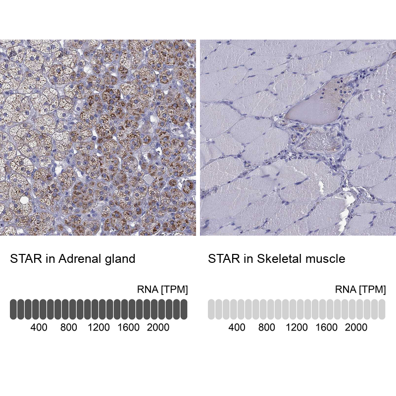
- Polyclonal Antibody against Human STAR, Gene description: steroidogenic acute regulatory protein, Alternative Gene Names: StAR, STARD1, Validated applications: IHC, Uniprot ID: P49675, Storage: Store at +4°C for short term storage. Long time storage is recommended at -20°C.
- Reactivity
- Human
- Host
- Rabbit
- Conjugate
- Unconjugated
- Isotype
- IgG
- Vial size
- 100 µl
- Concentration
- 0.1 mg/ml
- Storage
- Store at +4°C for short term storage. Long time storage is recommended at -20°C.
- Handling
- The antibody solution should be gently mixed before use.
Submitted references Incomplete Pattern of Steroidogenic Protein Expression in Functioning Adrenocortical Carcinomas.
The emerging role of the molecular marker p27 in the differential diagnosis of adrenocortical tumors.
Pereira SS, Costa MM, Gomez-Sanchez CE, Monteiro MP, Pignatelli D
Biomedicines 2020 Jul 30;8(8)
Biomedicines 2020 Jul 30;8(8)
The emerging role of the molecular marker p27 in the differential diagnosis of adrenocortical tumors.
Pereira SS, Morais T, Costa MM, Monteiro MP, Pignatelli D
Endocrine connections 2013;2(3):137-45
Endocrine connections 2013;2(3):137-45
No comments: Submit comment
Supportive validation
- Submitted by
- Atlas Antibodies (provider)
- Enhanced method
- Orthogonal validation
- Main image
- Experimental details
- Immunohistochemistry analysis in human adrenal gland and skeletal muscle tissues using HPA023644 antibody. Corresponding STAR RNA-seq data are presented for the same tissues.
- Sample type
- Human
- Protocol
- Protocol