Explore
 Validate
 Learn
LearnMAB1750-100
antibody from Novus Biologicals
Targeting: HAVCR1
CD365, HAVCR, HAVCR-1, KIM1, TIM-1, TIM1, TIMD1
 Western blot
Antibody data
- Antibody Data
- Antigen structure
- References [0]
- Comments [0]
- Validations
- Western blot [1]
- Immunohistochemistry [1]
- Flow cytometry [1]
Submit
Validation data
Reference
Comment
Report error
- Product number
- MAB1750-100 - Provider product page

- Provider
- Novus Biologicals
- Product name
- Mouse Monoclonal TIM-1/KIM-1/HAVCR Antibody
- Antibody type
- Monoclonal
- Description
- Protein A or G purified from hybridoma culture supernatant. Detects human TIM-1/KIM-1/HAVCR in direct ELISAs and Western blots. Does not cross-react with recombinant mouse (rm) TIM-1, rmTIM-2, or rhTIM-3.
- Reactivity
- Human
- Host
- Mouse
- Conjugate
- Unconjugated
- Isotype
- IgG
- Vial size
- 100 ug
- Concentration
- LYOPH
- Storage
- Use a manual defrost freezer and avoid repeated freeze-thaw cycles. 12 months from date of receipt, -20 to -70 degreesC as supplied. 1 month, 2 to 8 degreesC under sterile conditions after reconstitution. 6 months, -20 to -70 degreesC under sterile conditions after reconstitution.
No comments: Submit comment
Supportive validation
- Submitted by
- Novus Biologicals (provider)
- Main image
- Experimental details
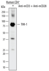
- Detection of Human TIM-1/KIM-1/HAVCR by Western Blot. Western blot shows lysates of human CD4+ cells treated (+) with 5 µg/mL of Hamster Anti-Mouse CD3 epsilon Monoclonal Antibody (Catalog # MAB484) and 1 µg/mL of Rat Anti-Mouse CD28 Monoclonal Antibody (Catalog # MAB4831) for 24 hours. PVDF membrane was probed with 1 µg/mL of Mouse Anti-Human TIM-1/KIM-1/HAVCR Monoclonal Antibody (Catalog # MAB1750) followed by HRP-conjugated Anti-Mouse IgG Secondary Antibody (Catalog # HAF007). A specific band was detected for TIM-1/KIM-1/HAVCR at approximately 80 kDa (as indicated). This experiment was conducted under reducing conditions and using Immunoblot Buffer Group 1.

Supportive validation
- Submitted by
- Novus Biologicals (provider)
- Main image

- Experimental details
- TIM-1/KIM-1/HAVCR in Human Kidney. TIM-1/KIM-1/HAVCR was detected in immersion fixed paraffin-embedded sections of human kidney using 25 µg/mL Mouse Anti-Human TIM-1/ KIM-1/HAVCR Monoclonal Antibody (Catalog # MAB1750) overnight at 4 °C. Tissue was stained with the Anti-Mouse HRP-DAB Cell & Tissue Staining Kit (brown; Catalog # CTS002) and counterstained with hematoxylin (blue). View our protocol for Chromogenic IHC Staining of Paraffin-embedded Tissue Sections.

Supportive validation
- Submitted by
- Novus Biologicals (provider)
- Main image

- Experimental details
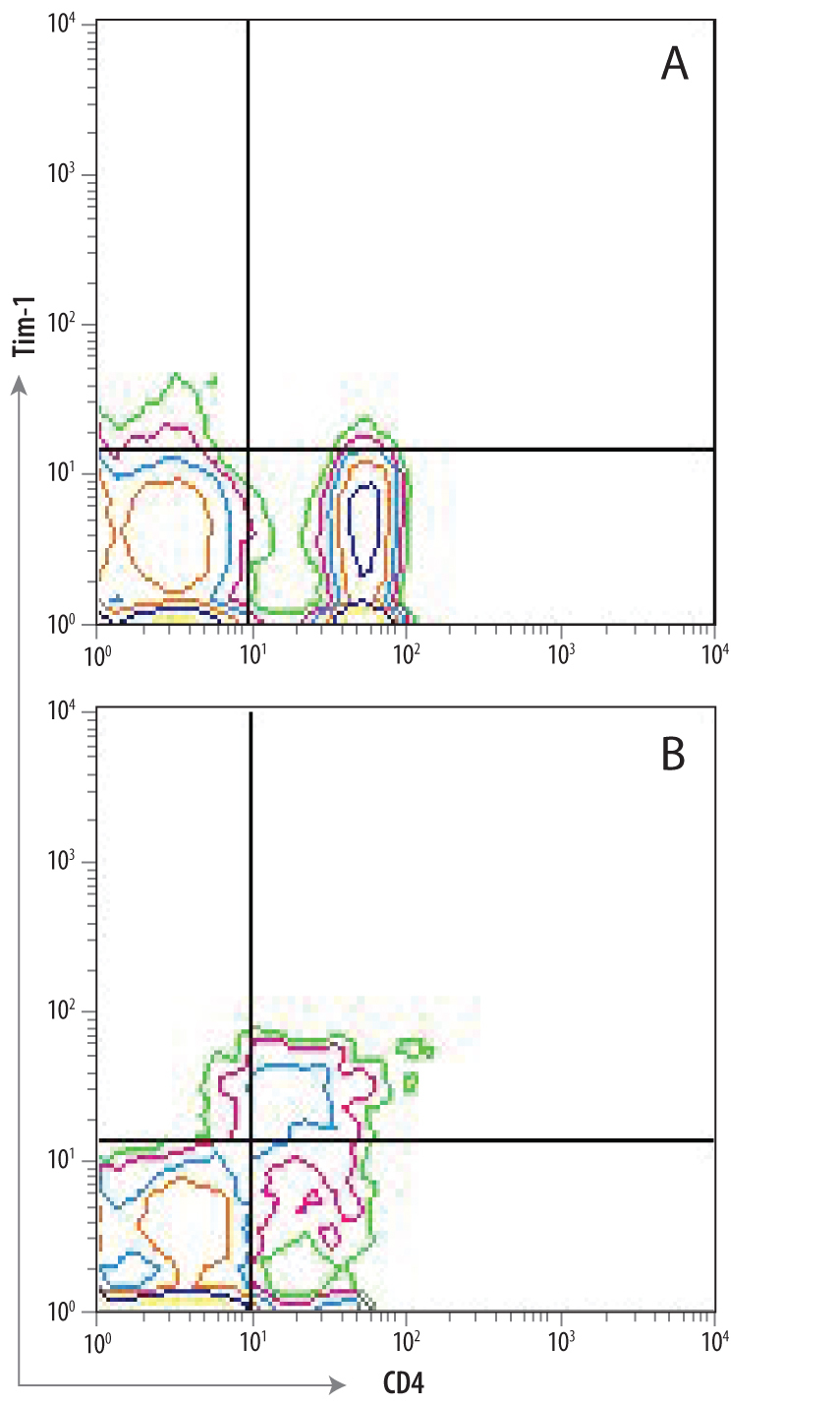
- Detection of TIM-1/KIM-1/HAVCR in Th2-stimulated Human PBMCs by Flow Cytometry. (A) Unstimulated and (B) Th2-stimulated human PBMCs were stained with Mouse Anti-Human TIM-1/KIM-1/HAVCR Monoclonal Antibody (Catalog # MAB1750) followed by Allophycocyanin-conjugated Anti-Mouse IgG Secondary Antibody (Catalog # F0101B) and Human CD4 PerCP-conjugated Monoclonal Antibody (Catalog # FAB3791C). Quadrant markers were set based on control antibody staining (Catalog # MAB0041).