Explore
 Validate
 Learn
LearnPA5-79345
antibody from Invitrogen Antibodies
Targeting: HAVCR1
CD365, HAVCR, HAVCR-1, KIM1, TIM-1, TIM1, TIMD1
 Western blot
 Immunocytochemistry
Antibody data
- Antibody Data
- Antigen structure
- References [3]
- Comments [0]
- Validations
- Immunocytochemistry [3]
- Immunohistochemistry [1]
- Other assay [2]
Submit
Validation data
Reference
Comment
Report error
- Product number
- PA5-79345 - Provider product page

- Provider
- Invitrogen Antibodies
- Product name
- KIM-1 Polyclonal Antibody
- Antibody type
- Polyclonal
- Antigen
- Synthetic peptide
- Description
- Reconstitute with 0.2 mL of distilled water to yield a concentration of 500 µg/mL. Positive Control - WB: SMMC Cell, HELA Cell, PANC Cell, M231 Cell, M453 Cell. IHC: Human Tonsil Tissue. ICC/IF: K562 Cell.
- Reactivity
- Human
- Host
- Rabbit
- Isotype
- IgG
- Vial size
- 100 μg
- Concentration
- 500 μg/mL
- Storage
- -20°C
Submitted references Deep Learning-Assisted Nephrotoxicity Testing with Bioprinted Renal Spheroids.
Paeonol Protects Against Methotrexate-Induced Nephrotoxicity via Upregulation of P-gp Expression and Inhibition of TLR4/NF-κB Pathway.
Quercetin improves contrast-induced acute kidney injury through the HIF-1α/lncRNA NEAT1/HMGB1 pathway.
Tröndle K, Miotto G, Rizzo L, Pichler R, Koch F, Koltay P, Zengerle R, Lienkamp SS, Kartmann S, Zimmermann S
International journal of bioprinting 2022;8(2):528
International journal of bioprinting 2022;8(2):528
Paeonol Protects Against Methotrexate-Induced Nephrotoxicity via Upregulation of P-gp Expression and Inhibition of TLR4/NF-κB Pathway.
Morsy MA, El-Sheikh AAK, Abdel-Hafez SMN, Kandeel M, Abdel-Gaber SA
Frontiers in pharmacology 2022;13:774387
Frontiers in pharmacology 2022;13:774387
Quercetin improves contrast-induced acute kidney injury through the HIF-1α/lncRNA NEAT1/HMGB1 pathway.
Luo M, Liu Z, Hu Z, He Q
Pharmaceutical biology 2022 Dec;60(1):889-898
Pharmaceutical biology 2022 Dec;60(1):889-898
No comments: Submit comment
Supportive validation
- Submitted by
- Invitrogen Antibodies (provider)
- Main image
- Experimental details
- Immunocytochemistry analysis of KIM-1 in K562 cells. Sample was incubated with KIM-1 polyclonal antibody (Product # PA5-79345).
- Submitted by
- Invitrogen Antibodies (provider)
- Main image
- Experimental details
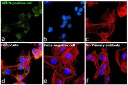
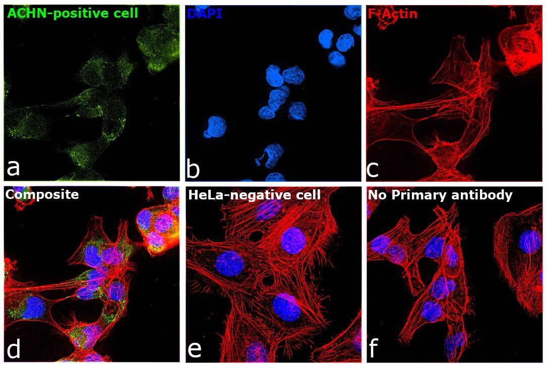
- Immunofluorescence analysis of KIM-1 was performed using 70% confluent log phase ACHN cells. The cells were fixed with 4% paraformaldehyde for 10 minutes, permeabilized with 0.1% Triton™ X-100 for 10 minutes, and blocked with 2% BSA for 45 minutes at room temperature. The cells were labeled with KIM-1 Polyclonal Antibody (Product # PA5-79345) at 1:200 dilution in 0.1% BSA, incubated at 4 degree celsius overnight and then labeled with Donkey anti-Rabbit IgG (H+L) Highly Cross-Adsorbed Secondary Antibody, Alexa Fluor Plus 488 (Product # A32790), (1:2500 dilution), for 45 minutes at room temperature (Panel a: Green). Nuclei (Panel b: Blue) were stained with ProLong™ Diamond Antifade Mountant with DAPI (Product # P36962). F-actin (Panel c: Red) was stained with Rhodamine Phalloidin (Product # R415, 1:300). Panel d represents the merged image showing Cytosolic, Golgi localization. Panel e represents negative expression model HeLa. Panel f represents control cells with no primary antibody to assess background. The images were captured at 60X magnification.
- Submitted by
- Invitrogen Antibodies (provider)
- Main image

- Experimental details
- Immunofluorescence analysis of KIM-1 was performed using 70% confluent log phase ACHN cells. The cells were fixed with 4% paraformaldehyde for 10 minutes, permeabilized with 0.1% Triton™ X-100 for 10 minutes, and blocked with 2% BSA for 45 minutes at room temperature. The cells were labeled with KIM-1 Polyclonal Antibody (Product # PA5-79345) at 1:200 dilution in 0.1% BSA, incubated at 4 degree celsius overnight and then labeled with Donkey anti-Rabbit IgG (H+L) Highly Cross-Adsorbed Secondary Antibody, Alexa Fluor Plus 488 (Product # A32790), (1:2500 dilution), for 45 minutes at room temperature (Panel a: Green). Nuclei (Panel b: Blue) were stained with ProLong™ Diamond Antifade Mountant with DAPI (Product # P36962). F-actin (Panel c: Red) was stained with Rhodamine Phalloidin (Product # R415, 1:300). Panel d represents the merged image showing Cytosolic, Golgi localization. Panel e represents negative expression model HeLa. Panel f represents control cells with no primary antibody to assess background. The images were captured at 60X magnification.

Supportive validation
- Submitted by
- Invitrogen Antibodies (provider)
- Main image
- Experimental details
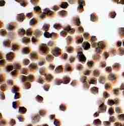
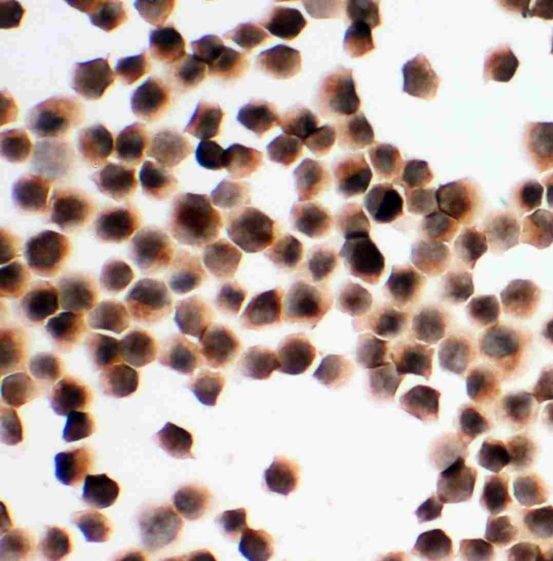
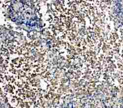
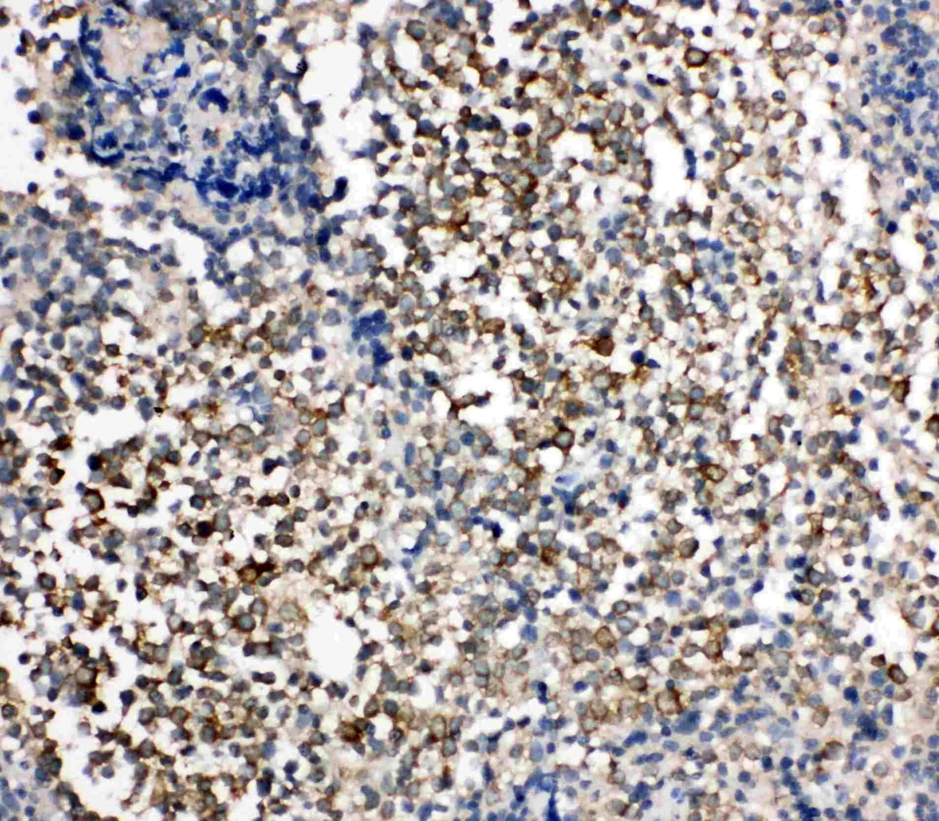
- Immunohistochemistry analysis of KIM-1 on paraffin-embedded human tonsil tissue. Sample was incubated with KIM-1 polyclonal antibody (Product# PA5-79345).
Supportive validation
- Submitted by
- Invitrogen Antibodies (provider)
- Main image
- Experimental details
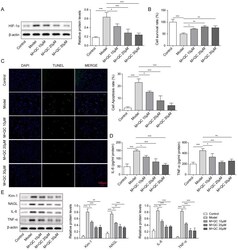
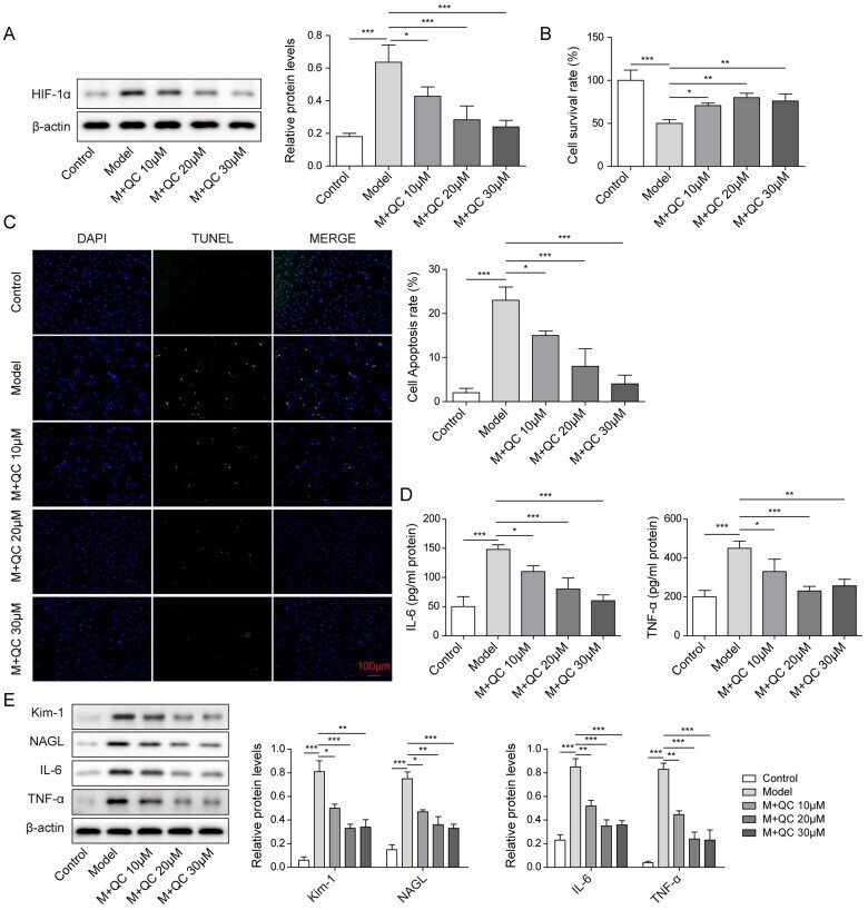
- Figure 2. Quercetin diminished cell injury and apoptosis in the CI-AKI model via inhibiting the expression of HIF-1alpha. (A) HIF-1alpha was assessed by western blotting in different groups treated with different concentrations of quercetin. (B) Cell survival rate was gradually increased in groups treated with 10, 20, and 30 muM quercetin. (C) Cell apoptosis rate was gradually decreased in model groups with quercetin concentration increased. (D) The expression levels of IL-6 and TNF-alpha were reduced with quercetin concentration increased in the CI-AKI model. (E) The expression levels of Kim-1, NAGL, IL-6 and TNF-alpha were decreased with the concentration of quercetin increased. The data were expressed as mean +- SD. * p < 0.05, ** p < 0.01.
- Submitted by
- Invitrogen Antibodies (provider)
- Main image
- Experimental details
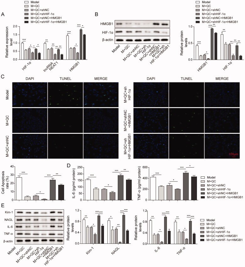
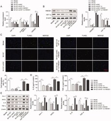
- Figure 5. Quercetin diminished cell injury and apoptosis in the CI-AKI model via inhibition of HIF-1alpha on the lncRNA NEAT1/HMGB1 signalling pathway. (A, B) Relative mRNA and protein expression level of HIF-1, lncRNA NEAT1 and HMGB1 in HK-2 cells of Model, M + QC, M + QC + sh NC, M + QC + sh HIF-1alpha, M + QC + sh NC + HMGB1, and M + QC + sh HIF-1alpha + HMGB1 group was measured by qRT-PCR and western blotting. HK-2 cells were subjected for the analysis of (C) cell apoptosis rate, (D) Elisa levels of IL-6 and TNF-alpha, (E) protein expression of IL-6, TNF-alpha, Kim-1, NAGL, IL-6 and TNF-alpha between M + QC and M + QC + sh-NC, M + QC + sh-NC and M + QC + sh-HIF-1, M + QC + sh-NC + HMGB1 and M + QC + sh-HIF-1alpha+ HMGB1 group. The data were expressed as mean +- SD. * p < 0.05, ** p < 0.01.