Explore
 Validate
 Learn
LearnPA1-41210
antibody from Invitrogen Antibodies
Targeting: HAVCR1
CD365, HAVCR, HAVCR-1, KIM1, TIM-1, TIM1, TIMD1
 Western blot
Antibody data
- Antibody Data
- Antigen structure
- References [0]
- Comments [0]
- Validations
- Western blot [2]
- Immunocytochemistry [2]
Submit
Validation data
Reference
Comment
Report error
- Product number
- PA1-41210 - Provider product page

- Provider
- Invitrogen Antibodies
- Product name
- TIM-1 Polyclonal Antibody
- Antibody type
- Polyclonal
- Antigen
- Synthetic peptide
- Description
- Suggested positive control: human and mouse brain tissue lysate or protein.
- Reactivity
- Human, Mouse
- Host
- Rabbit
- Isotype
- IgG
- Vial size
- 100 µg
- Concentration
- 1.0 mg/mL
- Storage
- Store at 4°C short term. For long term storage, store at -20°C, avoiding freeze/thaw cycles.
No comments: Submit comment
Supportive validation
- Submitted by
- Invitrogen Antibodies (provider)
- Main image

- Experimental details
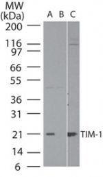
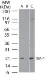
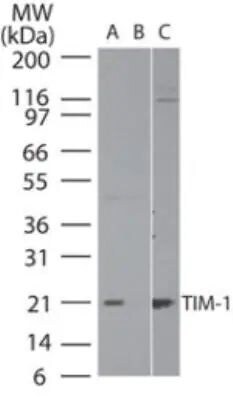
- Western blot analysis of TIM-1 in human brain lysatein the A) absence and B) presence of immunizing peptide, and C) mouse brain lysate using a Hepatitis A Virus Cellular Receptor 1 polyclonal antibody (Product # PA1-41210) at 2 µg/mL.
- Submitted by
- Invitrogen Antibodies (provider)
- Main image
- Experimental details
- Western blot analysis of TIM-1 in human brain lysate in the A) absence and B) presence of immunizing peptide, and C) mouse brain lysate. Samples were incubated in TIM-1 polyclonal antibody (Product # PA1-41210) using a dilution of 2 µg/mL.
Supportive validation
- Submitted by
- Invitrogen Antibodies (provider)
- Main image

- Experimental details
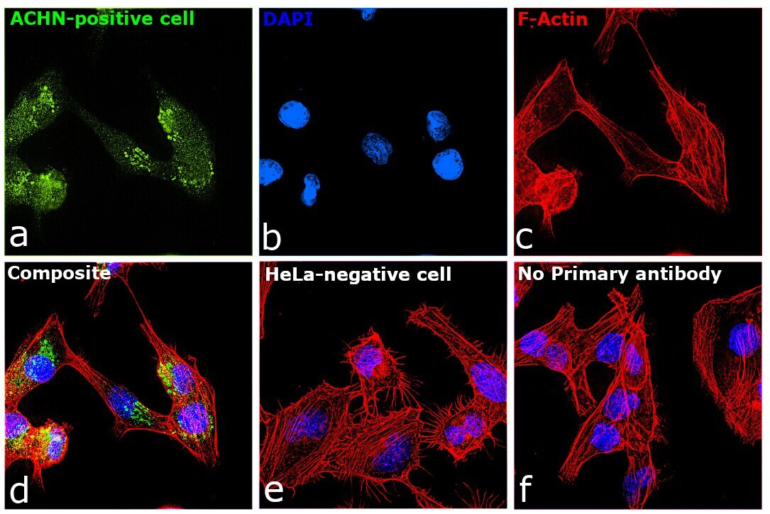
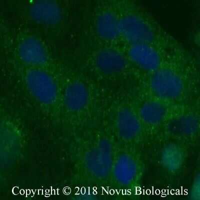
- Immunocytochemistry analysis of TIM-1 in Caco-2 cells fixed for 10 minutes using 10% formalin and then permeabilized for 5 minutes using 1X PBS + 0.05% Triton-X100. Samples were incubated in TIM-1 polyclonal antibody (Product # PA1-41210) using a dilution of 5 µg/mL overnight at 4 °C followed by anti-rabbit DyLight 488 (Green) with a dilution of 1:500. Nuclei were counterstained with DAPI (Blue). Cells were imaged using a 40X objective.
- Submitted by
- Invitrogen Antibodies (provider)
- Main image

- Experimental details
- Immunofluorescence analysis of TIM-1 was performed using 70% confluent log phase ACHN cells. The cells were fixed with 4% paraformaldehyde for 10 minutes, permeabilized with 0.1% Triton™ X-100 for 10 minutes, and blocked with 2% BSA for 45 minutes at room temperature. The cells were labeled with TIM-1 Polyclonal Antibody (Product # PA1-41210) at 1:200 dilution in 0.1% BSA, incubated at 4 degree celsius overnight and then labeled with Donkey anti-Rabbit IgG (H+L) Highly Cross-Adsorbed Secondary Antibody, Alexa Fluor Plus 488 (Product # A32790), (1:2500 dilution), for 45 minutes at room temperature (Panel a: Green). Nuclei (Panel b: Blue) were stained with ProLong™ Diamond Antifade Mountant with DAPI (Product # P36962). F-actin (Panel c: Red) was stained with Rhodamine Phalloidin (Product # R415, 1:300). Panel d represents the merged image showing Cytosolic, Golgi localization. Panel e represents negative expression model HeLa. Panel f represents control cells with no primary antibody to assess background. The images were captured at 60X magnification.