Explore
 Validate
 Learn
LearnPA5-20244
antibody from Invitrogen Antibodies
Targeting: HAVCR1
CD365, HAVCR, HAVCR-1, KIM1, TIM-1, TIM1, TIMD1
 Western blot
 Immunocytochemistry
Antibody data
- Antibody Data
- Antigen structure
- References [5]
- Comments [0]
- Validations
- Immunocytochemistry [5]
- Immunohistochemistry [1]
- Other assay [2]
Submit
Validation data
Reference
Comment
Report error
- Product number
- PA5-20244 - Provider product page

- Provider
- Invitrogen Antibodies
- Product name
- TIM-1 Polyclonal Antibody
- Antibody type
- Polyclonal
- Antigen
- Synthetic peptide
- Description
- A suggested positive control is human uterus tissue lysate. PA5-20244 can be used with blocking peptide PEP-0363. The PA5-20244 immunogen is located within amino acids 50 - 100 of TIM-1. Predicted molecular ~ 44kD. In Western blot applications, this antibody has been observed to detect a band at: 60kD (Post-modification: 4 N-linked glycosylation) Predicted species reactivity based on immunogen sequence: Mouse: (63%), Rat: (56%)
- Reactivity
- Human
- Host
- Rabbit
- Isotype
- IgG
- Vial size
- 100 μg
- Concentration
- 1 mg/mL
- Storage
- 4°C
Submitted references Exposure to Iron Oxide Nanoparticles Coated with Phospholipid-Based Polymeric Micelles Induces Renal Transitory Biochemical and Histopathological Changes in Mice.
Increased Endocytosis of Cadmium-Metallothionein through the 24p3 Receptor in an In Vivo Model with Reduced Proximal Tubular Activity.
The guanylate cyclase C agonist linaclotide ameliorates the gut-cardio-renal axis in an adenine-induced mouse model of chronic kidney disease.
Prognostic value of TIM-1 expression in human non-small-cell lung cancer.
Hydrogen sulfide inhibits Ca(2+)-induced mitochondrial permeability transition pore opening in type-1 diabetes.
Balas M, Popescu Din IM, Hermenean A, Cinteza LO, Dinischiotu A
Materials (Basel, Switzerland) 2021 May 17;14(10)
Materials (Basel, Switzerland) 2021 May 17;14(10)
Increased Endocytosis of Cadmium-Metallothionein through the 24p3 Receptor in an In Vivo Model with Reduced Proximal Tubular Activity.
Zavala-Guevara IP, Ortega-Romero MS, Narváez-Morales J, Jacobo-Estrada TL, Lee WK, Arreola-Mendoza L, Thévenod F, Barbier OC
International journal of molecular sciences 2021 Jul 6;22(14)
International journal of molecular sciences 2021 Jul 6;22(14)
The guanylate cyclase C agonist linaclotide ameliorates the gut-cardio-renal axis in an adenine-induced mouse model of chronic kidney disease.
Nanto-Hara F, Kanemitsu Y, Fukuda S, Kikuchi K, Asaji K, Saigusa D, Iwasaki T, Ho HJ, Mishima E, Suzuki T, Suzuki C, Tsukimi T, Matsuhashi T, Oikawa Y, Akiyama Y, Kure S, Owada Y, Tomioka Y, Soga T, Ito S, Abe T
Nephrology, dialysis, transplantation : official publication of the European Dialysis and Transplant Association - European Renal Association 2020 Feb 1;35(2):250-264
Nephrology, dialysis, transplantation : official publication of the European Dialysis and Transplant Association - European Renal Association 2020 Feb 1;35(2):250-264
Prognostic value of TIM-1 expression in human non-small-cell lung cancer.
Zheng X, Xu K, Chen L, Zhou Y, Jiang J
Journal of translational medicine 2019 May 28;17(1):178
Journal of translational medicine 2019 May 28;17(1):178
Hydrogen sulfide inhibits Ca(2+)-induced mitochondrial permeability transition pore opening in type-1 diabetes.
Papu John AS, Kundu S, Pushpakumar S, Amin M, Tyagi SC, Sen U
American journal of physiology. Endocrinology and metabolism 2019 Aug 1;317(2):E269-E283
American journal of physiology. Endocrinology and metabolism 2019 Aug 1;317(2):E269-E283
No comments: Submit comment
Supportive validation
- Submitted by
- Invitrogen Antibodies (provider)
- Main image
- Experimental details
- Immunofluorescent analysis of human uterus cells using a TIM-1 polyclonal antibody (Product # PA5-20244) at a 20 µg/mL dilution.
- Submitted by
- Invitrogen Antibodies (provider)
- Main image

- Experimental details
- Immunofluorescent analysis of 4% paraformaldehyde-fixed human uterus cells labeling TIM-1 with TIM-1 Polyclonal Antibody (Product # PA5-20244) at 20 µg/mL, followed by goat anti-rabbit IgG secondary antibody at 1:500 dilution (red). Image showing both membrane and cytoplasmic staining on human uterus cells.
- Submitted by
- Invitrogen Antibodies (provider)
- Main image

- Experimental details
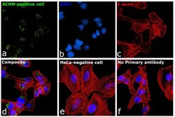
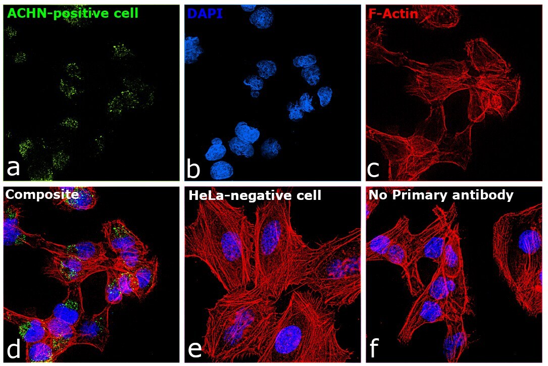
- Immunofluorescence analysis of TIM-1 was performed using 70% confluent log phase ACHN cells. The cells were fixed with 4% paraformaldehyde for 10 minutes, permeabilized with 0.1% Triton™ X-100 for 10 minutes, and blocked with 2% BSA for 45 minutes at room temperature. The cells were labeled with TIM-1 Polyclonal Antibody (Product # PA5-20244) at 1:200 dilution in 0.1% BSA, incubated at 4 degree celsius overnight and then labeled with Donkey anti-Rabbit IgG (H+L) Highly Cross-Adsorbed Secondary Antibody, Alexa Fluor Plus 488 (Product # A32790), (1:2500 dilution), for 45 minutes at room temperature (Panel a: Green). Nuclei (Panel b: Blue) were stained with ProLong™ Diamond Antifade Mountant with DAPI (Product # P36962). F-actin (Panel c: Red) was stained with Rhodamine Phalloidin (Product # R415, 1:300). Panel d represents the merged image showing Cytosolic, Golgi localization. Panel e represents negative expression model HeLa. Panel f represents control cells with no primary antibody to assess background. The images were captured at 60X magnification.

- Submitted by
- Invitrogen Antibodies (provider)
- Main image
- Experimental details
- Immunofluorescence analysis of TIM-1 was performed using 70% confluent log phase ACHN cells. The cells were fixed with 4% paraformaldehyde for 10 minutes, permeabilized with 0.1% Triton™ X-100 for 10 minutes, and blocked with 2% BSA for 45 minutes at room temperature. The cells were labeled with TIM-1 Polyclonal Antibody (Product # PA5-20244) at 1:200 dilution in 0.1% BSA, incubated at 4 degree celsius overnight and then labeled with Donkey anti-Rabbit IgG (H+L) Highly Cross-Adsorbed Secondary Antibody, Alexa Fluor Plus 488 (Product # A32790), (1:2500 dilution), for 45 minutes at room temperature (Panel a: Green). Nuclei (Panel b: Blue) were stained with ProLong™ Diamond Antifade Mountant with DAPI (Product # P36962). F-actin (Panel c: Red) was stained with Rhodamine Phalloidin (Product # R415, 1:300). Panel d represents the merged image showing Cytosolic, Golgi localization. Panel e represents negative expression model HeLa. Panel f represents control cells with no primary antibody to assess background. The images were captured at 60X magnification.
- Submitted by
- Invitrogen Antibodies (provider)
- Main image
- Experimental details
- Immunofluorescent analysis of 4% paraformaldehyde-fixed human uterus cells labeling TIM-1 with TIM-1 Polyclonal Antibody (Product # PA5-20244) at 20 µg/mL, followed by goat anti-rabbit IgG secondary antibody at 1:500 dilution (red). Image showing both membrane and cytoplasmic staining on human uterus cells.

Supportive validation
- Submitted by
- Invitrogen Antibodies (provider)
- Main image
- Experimental details
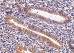
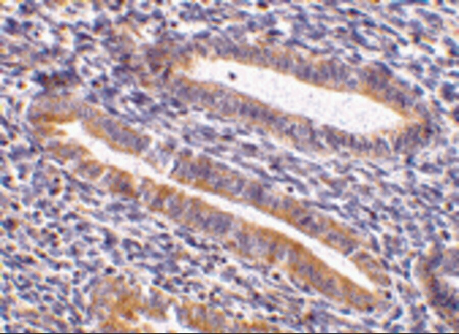
- Immunohistochemical analysis of paraffin-embedded human uterus tissue using TIM-1 Polyclonal Antibody (Product # PA5-20244) at 10 µg/mL. Tissue was fixed with formaldehyde and blocked with 0.1 serum for 1 h at RT; antigen retrieval was by heat mediation with a citrate buffer (pH6). Samples were incubated with primary antibody overnight at 4˚ C. A goat anti-rabbit IgG H&L (HRP) at 1/250 was used as secondary. Counter stained with Hematoxylin.
Supportive validation
- Submitted by
- Invitrogen Antibodies (provider)
- Main image
- Experimental details
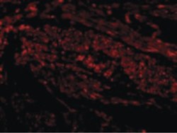
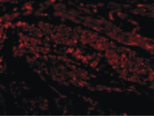
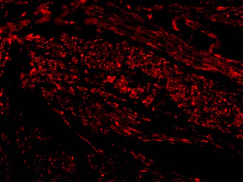
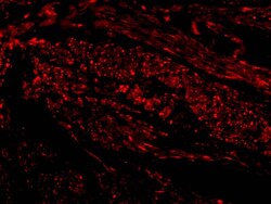
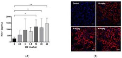
- Figure 2 Kim-1 protein increases after GM administration for 7 days. ( A ) Quantification of Kim-1 protein in whole renal tissue using xMAP technology. Bar graph shows means +- SD. n = 5 animals per dose. One-way ANOVA was performed p = 0.0071. Post-hoc: Dunn test. * p < 0.05; ** p < 0.005.; ( B ) Representative micrographs of the expression pattern of Kim-1 protein (red fluorescence) in renal tissue after the exposure to 10, 20 and 40 mg of GM/kg/day. The nuclei were stained with 4,6-diamino-2-phenylindole (DAPI, blue). Scale bars represent 50 um.

- Submitted by
- Invitrogen Antibodies (provider)
- Main image

- Experimental details
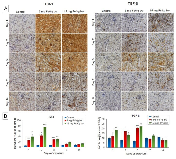
- Figure 3 The effect of IONPs encapsulated in phospholipid-based micelles on the TIM-1 and TGF-beta expression and distribution in the CD1 mice kidney tissue at 1, 2, 3, and 7 days post-exposure. ( A ) Immunohistochemistry (IHC) images; ( B ) quantification of IHC images. The IHC score was expressed in percentage of the stained area. The results are calculated as the mean +- standard deviation (SD) and are considered statistically significant when * p < 0.05; ** p < 0.01; *** p < 0.001 versus the control group. Scale bar: 20 mum.