 Explore
 Validate
 Learn
LearnPA5-20245
antibody from Invitrogen Antibodies
Targeting: HAVCR1
CD365, HAVCR, HAVCR-1, KIM1, TIM-1, TIM1, TIMD1
 Western blot
Antibody data
- Antibody Data
- Antigen structure
- References [0]
- Comments [0]
- Validations
- Western blot [3]
- Immunocytochemistry [1]
Submit
Validation data
Reference
Comment
Report error
- Product number
- PA5-20245 - Provider product page

- Provider
- Invitrogen Antibodies
- Product name
- TIM-1 Polyclonal Antibody
- Antibody type
- Polyclonal
- Antigen
- Synthetic peptide
- Description
- A suggested positive control is human uterus tissue lysate. PA5-20245 can be used with blocking peptide PEP-0364. The PA5-20245 immunogen is located within amino acids 230 - 280 of TIM-1. Predicted molecular ~ 44kD. In Western blot applications, this antibody has been observed to detect a band at: 60kD (Post-modification: 4 N-linked glycosylation) Predicted species reactivity based on immunogen sequence: Mouse: (50%), Rat: (44%)
- Reactivity
- Human
- Host
- Rabbit
- Isotype
- IgG
- Vial size
- 100 µg
- Concentration
- 1 mg/mL
- Storage
- Maintain refrigerated at 2-8°C for up to 3 months. For long term storage store at -20°C
No comments: Submit comment
Supportive validation
- Submitted by
- Invitrogen Antibodies (provider)
- Main image
- Experimental details
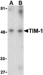
- Western blot analysis of human uterus tissue lysate using a TIM-1 polyclonal antibody (Product # PA5-20245) at (A) 1 and (B) 2 µg/mL.

- Submitted by
- Invitrogen Antibodies (provider)
- Main image
- Experimental details
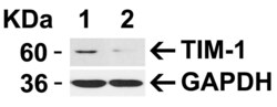
- Western Bloat analysis TIM-1 siRNA Knockdown in HeLa Cells. HeLa cells were transfected with control siRNAs (lane 1) or TIM-1 siRNAs (lane 2) Loading: 15 µg of HeLa whole cell lysates per lane. Antibodies: TIM-1 Polyclonal Antibody (Product # PA5-20245) (1 µg/mL), 1 h incubation at RT in 5% NFDM/TBST. Secondary: Goat anti-rabbit IgG HRP conjugate at 1:10,000 dilution.

- Submitted by
- Invitrogen Antibodies (provider)
- Main image
- Experimental details
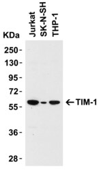
- Western Blot Validation in Human Cell Lines. Loading: 15 µg of lysates per lane. Antibodies: TIM-1 Polyclonal Antibody (Product # PA5-20245) (1 µg/mL), 1 h incubation at RT in 0.05 NFDM/TBST. Secondary: Goat anti-rabbit IgG HRP conjugate at 1:10,000 dilution.

Supportive validation
- Submitted by
- Invitrogen Antibodies (provider)
- Main image

- Experimental details
- Immunofluorescence analysis of TIM-1 was performed using 70% confluent log phase ACHN cells. The cells were fixed with 4% paraformaldehyde for 10 minutes, permeabilized with 0.1% Triton™ X-100 for 10 minutes, and blocked with 2% BSA for 45 minutes at room temperature. The cells were labeled with TIM-1 Polyclonal Antibody (Product # PA5-20245) at 1:200 dilution in 0.1% BSA, incubated at 4 degree celsius overnight and then labeled with Donkey anti-Rabbit IgG (H+L) Highly Cross-Adsorbed Secondary Antibody, Alexa Fluor Plus 488 (Product # A32790), (1:2500 dilution), for 45 minutes at room temperature (Panel a: Green). Nuclei (Panel b: Blue) were stained with ProLong™ Diamond Antifade Mountant with DAPI (Product # P36962). F-actin (Panel c: Red) was stained with Rhodamine Phalloidin (Product # R415, 1:300). Panel d represents the merged image showing Cytosolic, Golgi localization. Panel e represents negative expression model HeLa. Panel f represents control cells with no primary antibody to assess background. The images were captured at 60X magnification.
