Explore
 Validate
 Learn
Learn Western blot
 Immunohistochemistry
Antibody data
- Antibody Data
- Antigen structure
- References [7]
- Comments [0]
- Validations
- Immunohistochemistry [1]
Submit
Validation data
Reference
Comment
Report error
- Product number
- AF1750 - Provider product page

- Provider
- R&D Systems
- Product name
- Human TIM-1/KIM-1/HAVCR Antibody
- Antibody type
- Polyclonal
- Description
- Antigen Affinity-purified. Detects human TIM-1/KIM-1/HAVCR in ELISAs and Western blots. In sandwich immunoassays, less than 0.1% cross-reactivity with recombinant mouse (rm) TIM-1, recombinant rat TIM-1, and recombinant human (rh) TIM-4 is observed.
- Reactivity
- Human
- Host
- Goat
- Conjugate
- Unconjugated
- Antigen sequence
Q96D42- Isotype
- IgG
- Vial size
- 100 ug
- Concentration
- LYOPH
- Storage
- Use a manual defrost freezer and avoid repeated freeze-thaw cycles. 12 months from date of receipt, -20 to -70 °C as supplied. 1 month, 2 to 8 °C under sterile conditions after reconstitution. 6 months, -20 to -70 °C under sterile conditions after reconstitution.
Submitted references Dynamic Dystroglycan Complexes Mediate Cell Entry of Lassa Virus.
TIM-1 Promotes Japanese Encephalitis Virus Entry and Infection.
Human Sertoli cells support high levels of Zika virus replication and persistence.
Urinary kidney injury molecule‑1 as an early diagnostic biomarker of obstructive acute kidney injury and development of a rapid detection method.
Generation of nephron progenitor cells and kidney organoids from human pluripotent stem cells.
Nanoparticle Detection of Urinary Markers for Point-of-Care Diagnosis of Kidney Injury.
Characterizing functional domains for TIM-mediated enveloped virus entry.
Herrador A, Fedeli C, Radulovic E, Campbell KP, Moreno H, Gerold G, Kunz S
mBio 2019 Mar 26;10(2)
mBio 2019 Mar 26;10(2)
TIM-1 Promotes Japanese Encephalitis Virus Entry and Infection.
Niu J, Jiang Y, Xu H, Zhao C, Zhou G, Chen P, Cao R
Viruses 2018 Nov 14;10(11)
Viruses 2018 Nov 14;10(11)
Human Sertoli cells support high levels of Zika virus replication and persistence.
Kumar A, Jovel J, Lopez-Orozco J, Limonta D, Airo AM, Hou S, Stryapunina I, Fibke C, Moore RB, Hobman TC
Scientific reports 2018 Apr 3;8(1):5477
Scientific reports 2018 Apr 3;8(1):5477
Urinary kidney injury molecule‑1 as an early diagnostic biomarker of obstructive acute kidney injury and development of a rapid detection method.
Jin Y, Shao X, Sun B, Miao C, Li Z, Shi Y
Molecular medicine reports 2017 Mar;15(3):1229-1235
Molecular medicine reports 2017 Mar;15(3):1229-1235
Generation of nephron progenitor cells and kidney organoids from human pluripotent stem cells.
Morizane R, Bonventre JV
Nature protocols 2017 Jan;12(1):195-207
Nature protocols 2017 Jan;12(1):195-207
Nanoparticle Detection of Urinary Markers for Point-of-Care Diagnosis of Kidney Injury.
Chung HJ, Pellegrini KL, Chung J, Wanigasuriya K, Jayawardene I, Lee K, Lee H, Vaidya VS, Weissleder R
PloS one 2015;10(7):e0133417
PloS one 2015;10(7):e0133417
Characterizing functional domains for TIM-mediated enveloped virus entry.
Moller-Tank S, Albritton LM, Rennert PD, Maury W
Journal of virology 2014 Jun;88(12):6702-13
Journal of virology 2014 Jun;88(12):6702-13
No comments: Submit comment
Supportive validation
- Submitted by
- R&D Systems (provider)
- Main image

- Experimental details
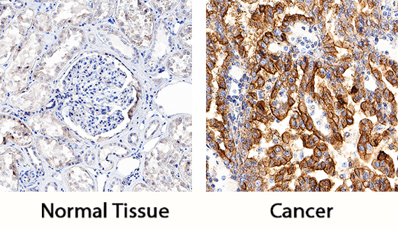
- TIM-1/KIM-1/HAVCR in Human Kidney Cancer Tissue. TIM-1/KIM-1/HAVCR was detected in immersion fixed paraffin-embedded sections of human kidney cancer tissue using Goat Anti-Human TIM-1/KIM-1/HAVCR Antigen Affinity-purified Polyclonal Antibody (Catalog # AF1750) at 1 µg/mL for 1 hour at room temperature followed by incubation with the Anti-Goat IgG VisUCyte™ HRP Polymer Antibody (Catalog # VC004). Tissue was stained using DAB (brown) and counterstained with hematoxylin (blue). Specific staining was localized to cancer cells. View our protocol for IHC Staining with VisUCyte HRP Polymer Detection Reagents.